Copyright
©The Author(s) 2025.
World J Gastrointest Surg. Jun 27, 2025; 17(6): 107086
Published online Jun 27, 2025. doi: 10.4240/wjgs.v17.i6.107086
Published online Jun 27, 2025. doi: 10.4240/wjgs.v17.i6.107086
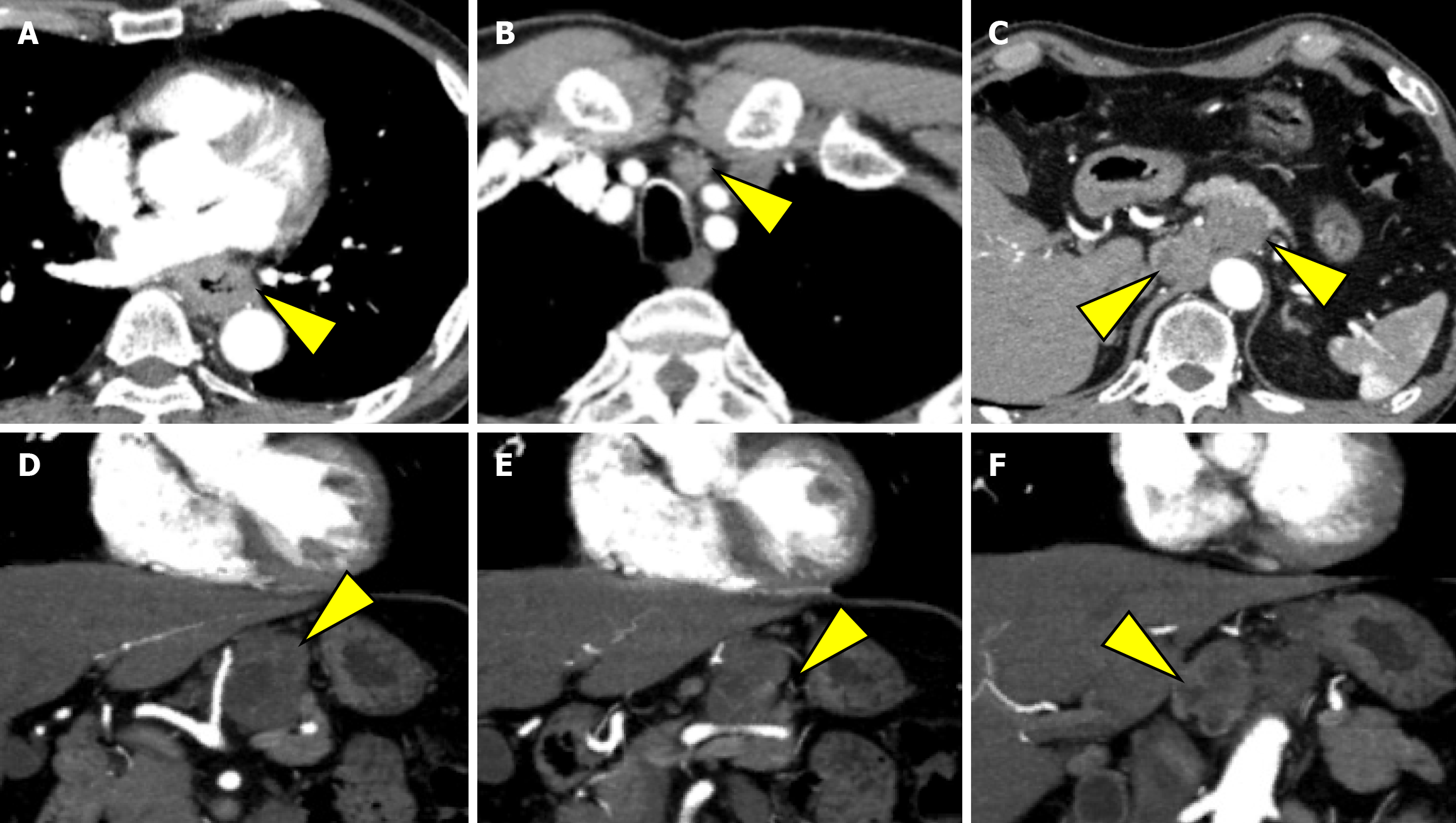
Figure 2 Computed tomography findings at the initial presentation.
A: Image showing irregular wall thickening from the middle to the lower thoracic esophagus as the primary lesion; B: Imaging showing enlarged No. 101L lymph node measuring 15 mm; C: Imaging showing bulky suprapancreatic lymph node metastasis, including No. 8, 9, and 11p; D and E: Imaging showing enlargement of the No. 11p lymph node to 35 mm, directly invading the pancreatic body; F: Imaging showing enlarged No. 8 and No. 9 lymph nodes, forming a mass.
- Citation: Okamoto K, Fujisawa K, Kono K, Ogawa Y, Shimoyama H, Haruta S, Takazawa Y, Ueno M, Udagawa H. Long-term survival with multimodal treatment including conversion surgery for locally advanced esophageal neuroendocrine carcinoma: A case report. World J Gastrointest Surg 2025; 17(6): 107086
- URL: https://www.wjgnet.com/1948-9366/full/v17/i6/107086.htm
- DOI: https://dx.doi.org/10.4240/wjgs.v17.i6.107086


