Copyright
©The Author(s) 2025.
World J Stem Cells. May 26, 2025; 17(5): 105394
Published online May 26, 2025. doi: 10.4252/wjsc.v17.i5.105394
Published online May 26, 2025. doi: 10.4252/wjsc.v17.i5.105394
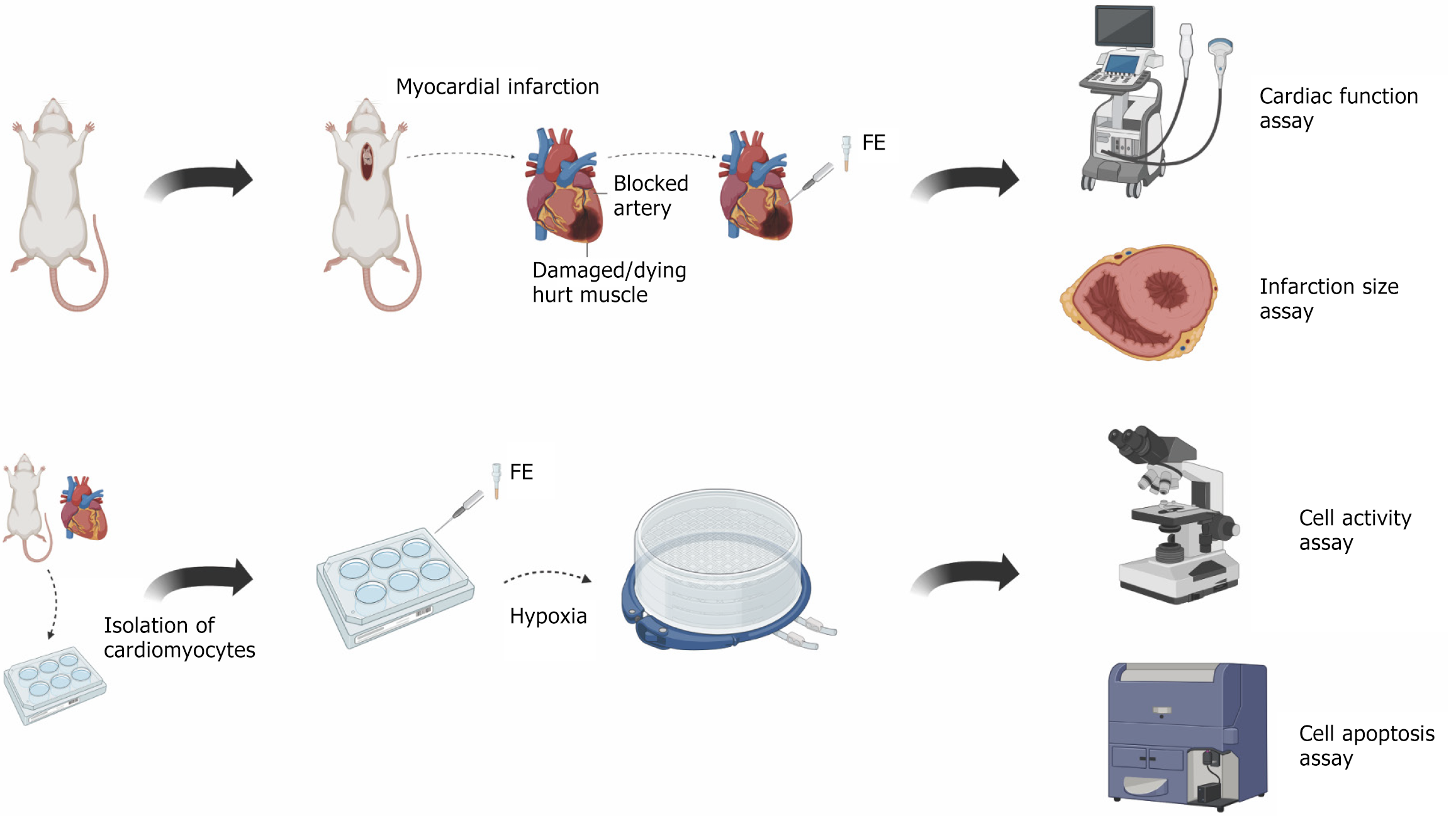
Figure 1 Schematic illustration of experimental process.
FE: Fat extract.
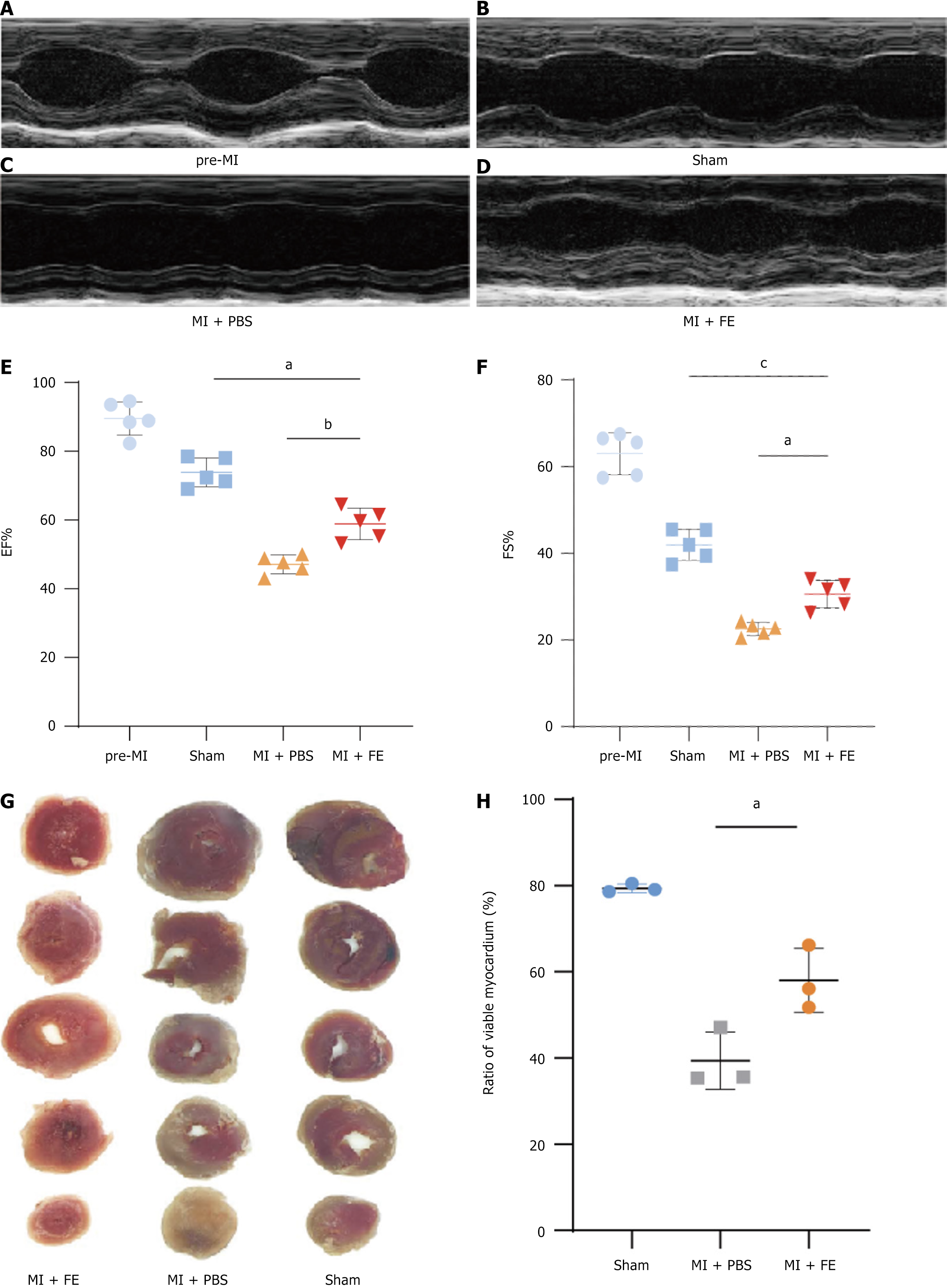
Figure 2 Fat extract injection improved cardiac function after myocardial infarction in mice.
A-D: Representative M-mode images of transthoracic echocardiography; E and F: Quantification of ejection fraction (EF) and fraction shortening (FS) (n = 5); G: Representative images of triphenyl-2H-tetrazolium chloride-staining (scale bar = 2 mm, n = 3); H: Quantification of infarct size. Values indicate mean values ± SEM from at least three independent experiments. aP < 0.05, bP < 0.01, cP < 0.001. MI: Myocardial infarction; PBS: Phosphate-buffered saline; FE: Fat extract; EF: Ejection fraction; FS: Fraction shortening.
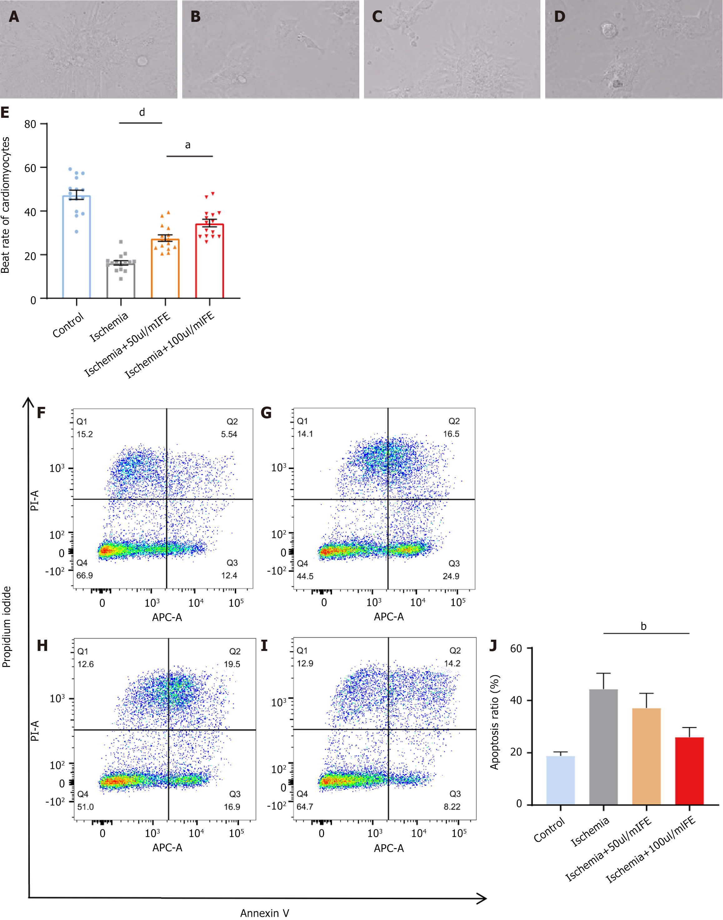
Figure 3 Cell viability of cardiomyocytes was higher and apoptosis level in cardiac myocytes was lower in the treatment than in the control group.
A-D: Beat rate of cardiomyocytes determined by live cell imaging (n = 15 cells per group); E: Quantification of beat rate of cardiomyocytes; F-I: The level of cell apoptosis were calculated using flow cytometry (n = 3); J: Quantification of cell apoptosis. Values indicate mean ± SEM from at least three independent experiments. aP < 0.05, bP < 0.01, dP < 0.0001. FE: Fat extract.
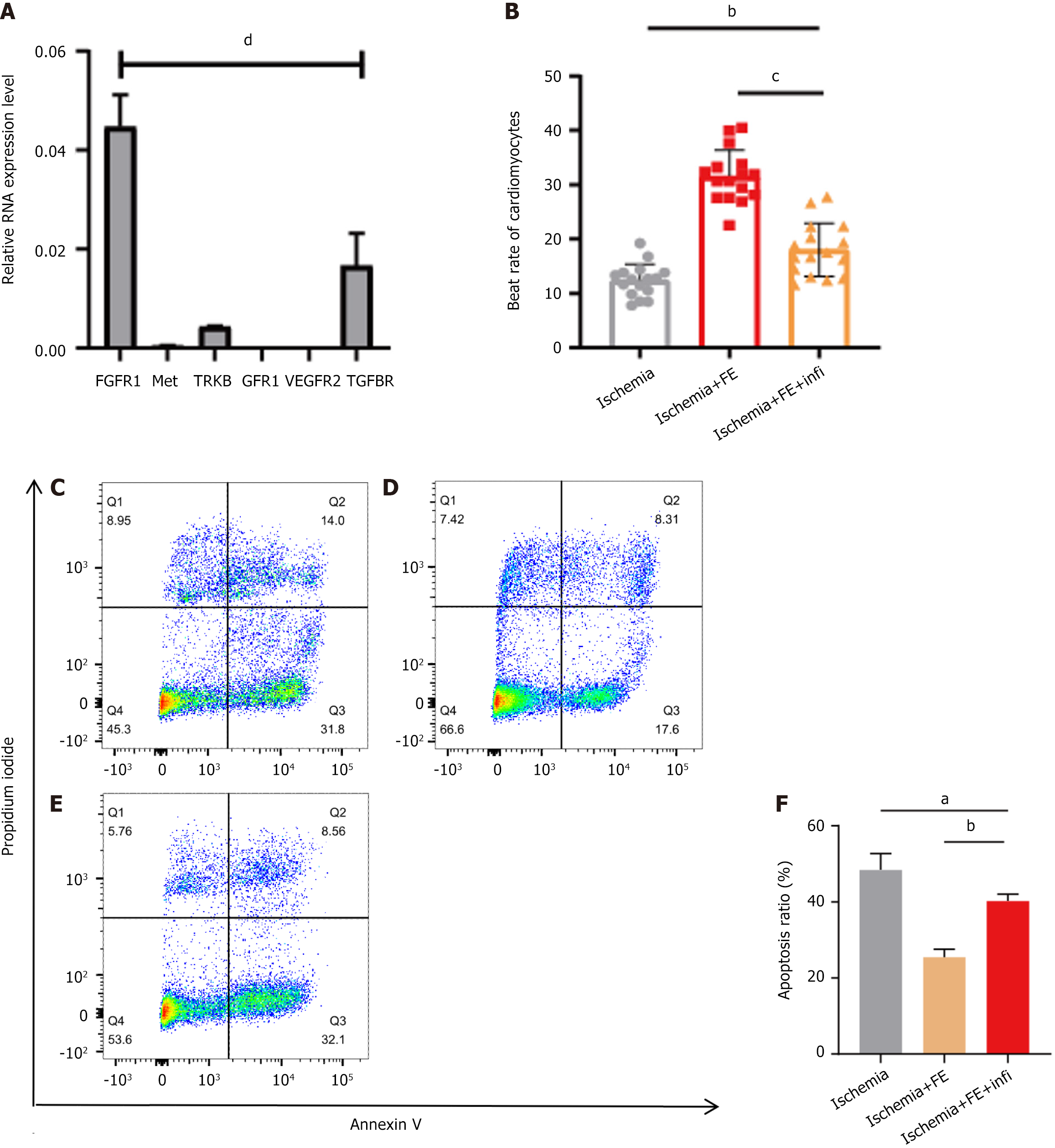
Figure 4 Fibroblast growth factor receptor family inhibitor partly blocked the therapeutic effect of fat extract at the cellular level after ischaemia injury.
A: Relative mRNA expression levels of tropomyosin receptor kinase B, transforming growth factor-beta receptor, Met, fibroblast growth factor receptor 1, vascular endothelial growth factor receptor 2, GFR1 in cardiomyocytes; B Quantification of beat rate of cardiomyocytes; C-E: The level of cell apoptosis were calculated using flow cytometry (n = 3); F: Quantification of cell apoptosis. Values indicate mean ± SEM from at least three independent experiments. aP < 0.05, bP < 0.01, cP < 0.001, dP < 0.0001. TRKB: Tropomyosin receptor kinase B; TGFBR: Transforming growth factor-beta receptor; FGFR1: Fibroblast growth factor receptor 1; VEGFR2: Vascular endothelial growth factor receptor 2; FE: Fat extract; Infi: Infigratinib.
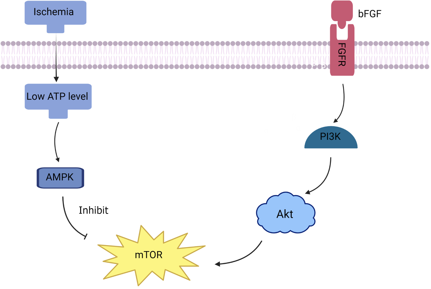
Figure 5 Basic fibroblast growth factor-phosphatidylinositol 3-kinase/protein kinase B/mechanistic target of rapamycin signaling pathways in ischemia condition.
bFGF: Basic fibroblast growth factor; FGFR: Fibroblast growth factor receptor; AMPK: AMP-activated protein kinase; mTOR: Mechanistic target of rapamycin; PI3K: Phosphatidylinositol 3-kinase; Akt: Protein kinase B.
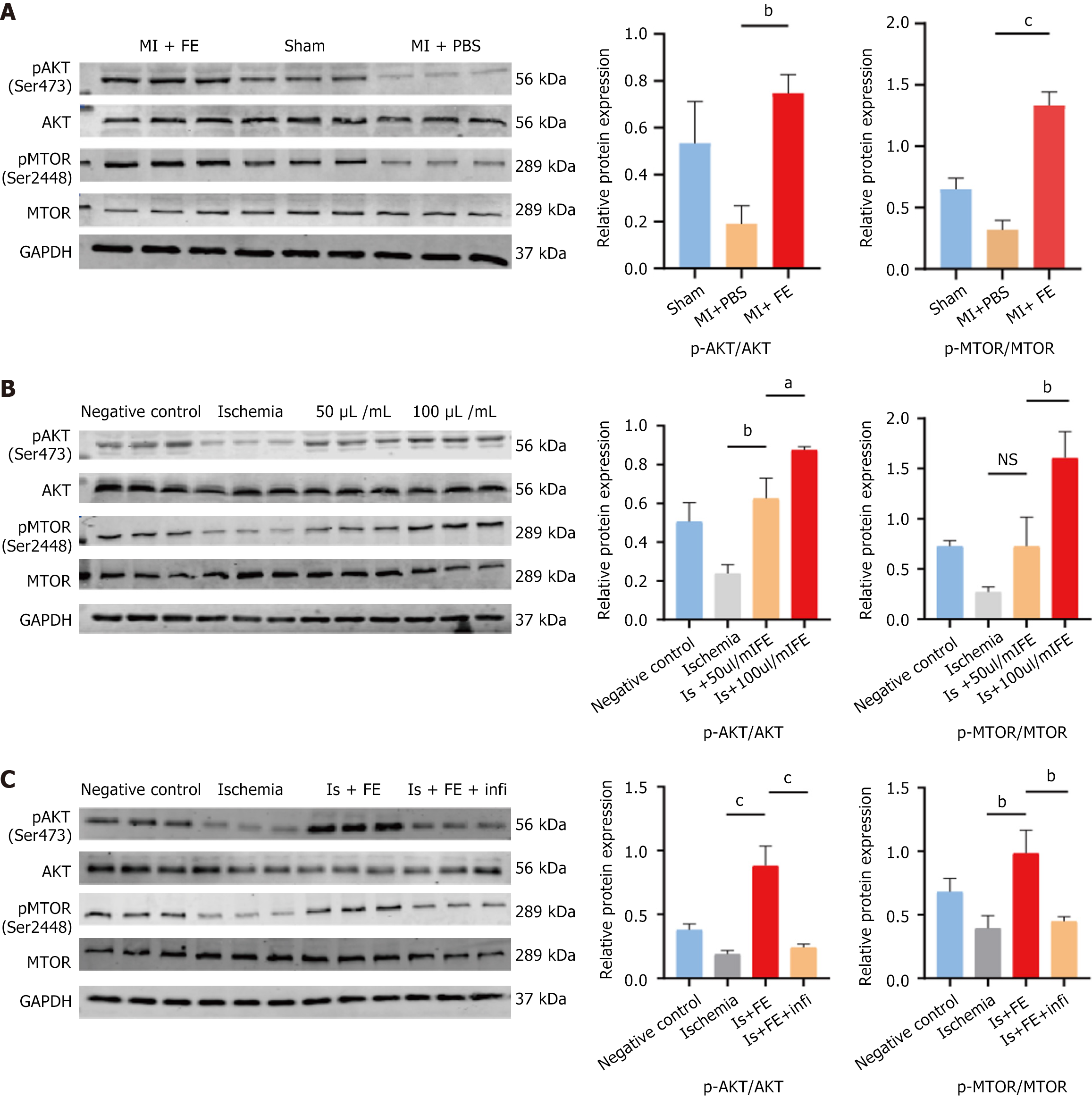
Figure 6 Activation of the basic fibroblast growth factor-phosphatidylinositol 3-kinase/protein kinase B/mechanistic target of rapamycin signaling pathways may be involved in the role of fat extract in myocardial ischemia injury.
The expression and phosphorylation levels of protein kinase B and mechanistic target of rapamycin were analyzed by western blot. A: C57 mice myocardial tissues; B and C: Hypoxia treated cardiomyocytes. Data were presented as mean ± SEM (n = 3 per group), aP < 0.05, bP < 0.01, cP < 0.001, NS: No significance. MI: Myocardial infarction; PBS: Phosphate-buffered saline; FE: Fat extract; Akt: Protein kinase B; mTOR: Mechanistic target of rapamycin.
- Citation: Yang TY, Sun Y, Zhang WJ, Wang CQ, Zhou J. Cell-free extracts from human fat tissue attenuate ischemic injury in cardiomyocytes in a murine model. World J Stem Cells 2025; 17(5): 105394
- URL: https://www.wjgnet.com/1948-0210/full/v17/i5/105394.htm
- DOI: https://dx.doi.org/10.4252/wjsc.v17.i5.105394


