
















Copyright: ©Author(s) 2026.
World J Gastroenterol. Nov 28, 2026; 32(44): 121025
Published online Nov 28, 2026. doi: 10.3748/wjg.121025
Published online Nov 28, 2026. doi: 10.3748/wjg.121025
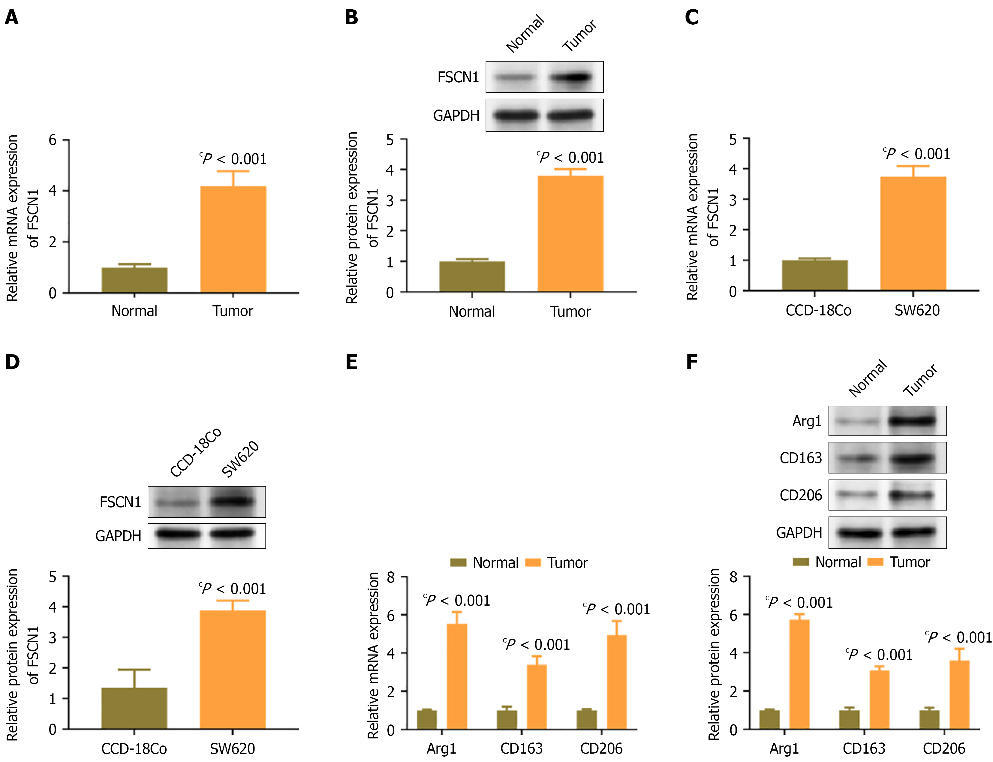
Figure 1 Fascin actin-bundling protein-1 is highly expressed in colon cancer tissues and cell lines.
A and B: The mRNA and protein expression levels of fascin actin-bundling protein-1 in colon cancer tissues; C and D: The mRNA and protein expression levels of fascin actin-bundling protein-1 in colon cancer cell line SW620 and normal colon epithelial cell line CCD-18Co; E and F: Expression levels of M2 macrophage markers arginase 1, CD163, and CD206 in colon cancer tissues and their paired adjacent tissues. cP < 0.001. FSCN1: Fascin actin-bundling protein-1; Arg1: Arginase 1.
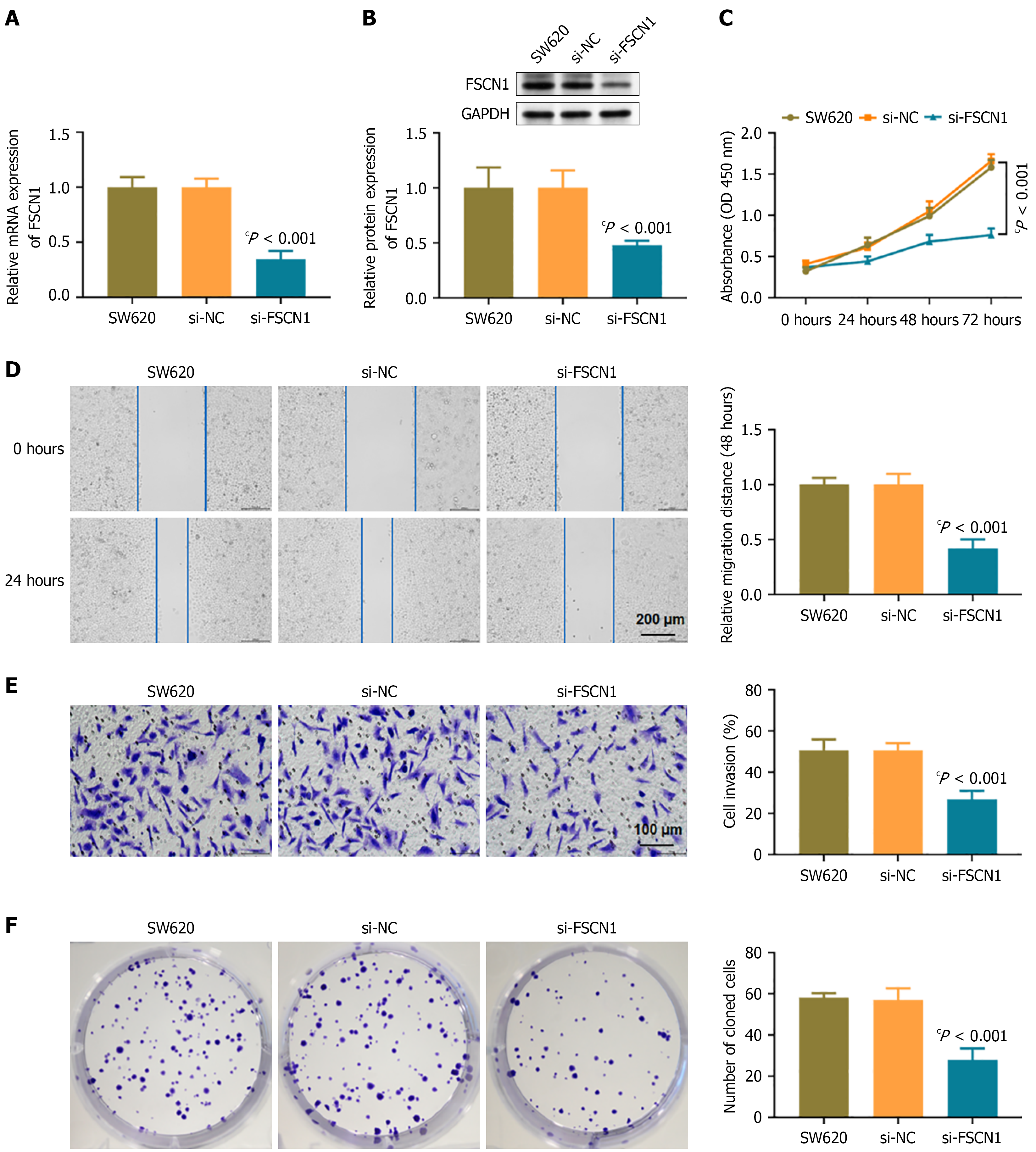
Figure 2 Fascin actin-bundling protein-1 silencing inhibits the malignant phenotype of SW620 cells.
A and B: The mRNA and protein expression levels of fascin actin-bundling protein-1; C: Cell proliferation; D and E: Transwell cell migration and cell invasion. Scale bar = 200 μm and 100 μm; F: Clone formation experiment. cP < 0.001. FSCN1: Fascin actin-bundling protein-1; NC: Non-targeting control.
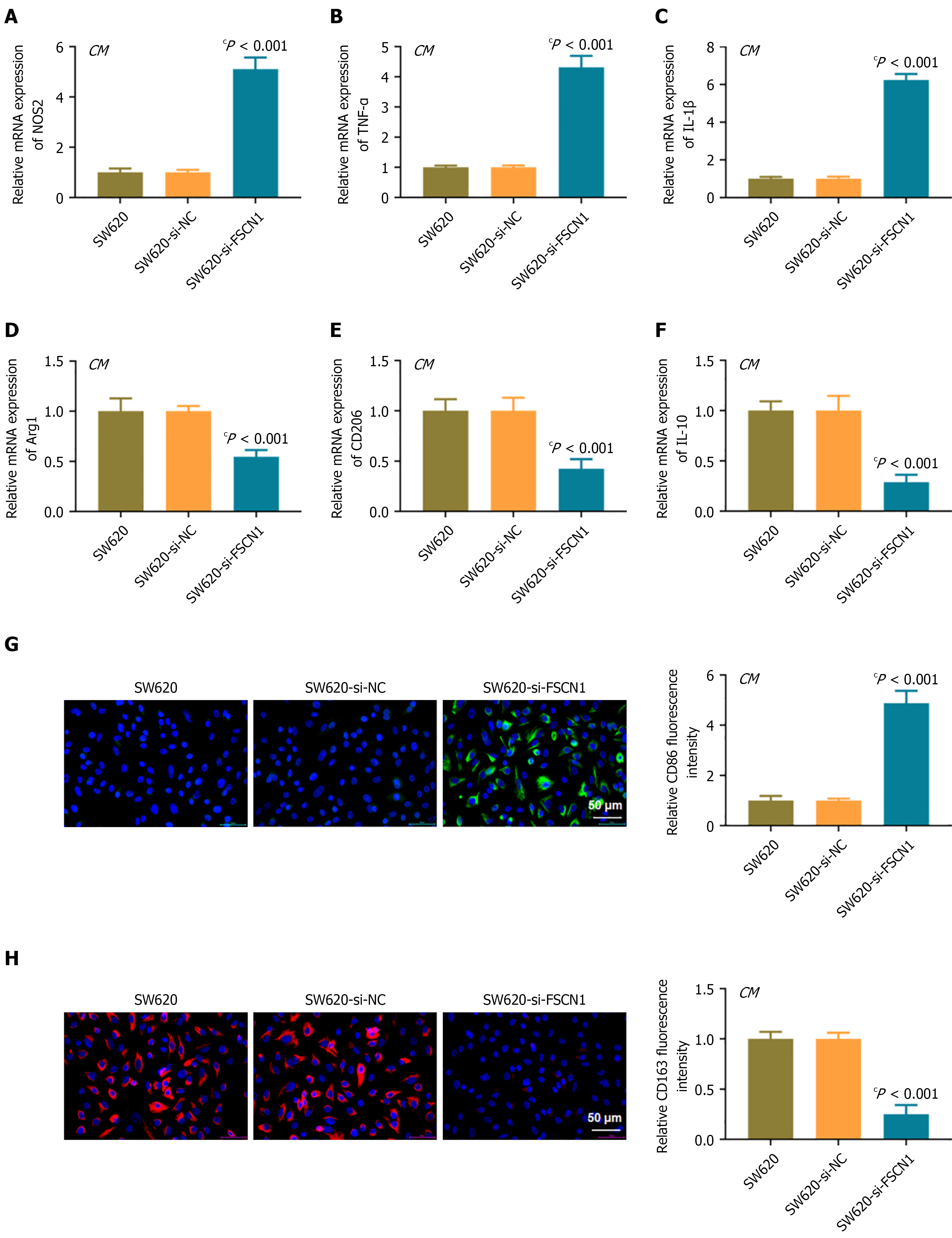
Figure 3 Knockdown of Fascin actin-bundling protein-1 inhibits M2 phenotype of macrophages.
A-C: The mRNA expression levels of M1 markers NOS2, tumor necrosis factor-α, and interleukin-1β; D-F: The mRNA expression levels of M2 markers arginase 1, CD206, and interleukin-10; G and H: Immunofluorescence staining of M1 marker CD86 and M2 marker CD163. Scale bar = 50 μm. cP < 0.001. THP-1 cells were induced to differentiate into macrophages by PMA and treated with conditioned medium containing SW620, SW620-si-NC, and SW620-si-FSCN1 cells, respectively. TNF-α: Tumor necrosis factor-α; FSCN1: Fascin actin-bundling protein-1; CM: Conditioned medium; Arg1: Arginase 1; IL: Interleukin; NC: Non-targeting control.
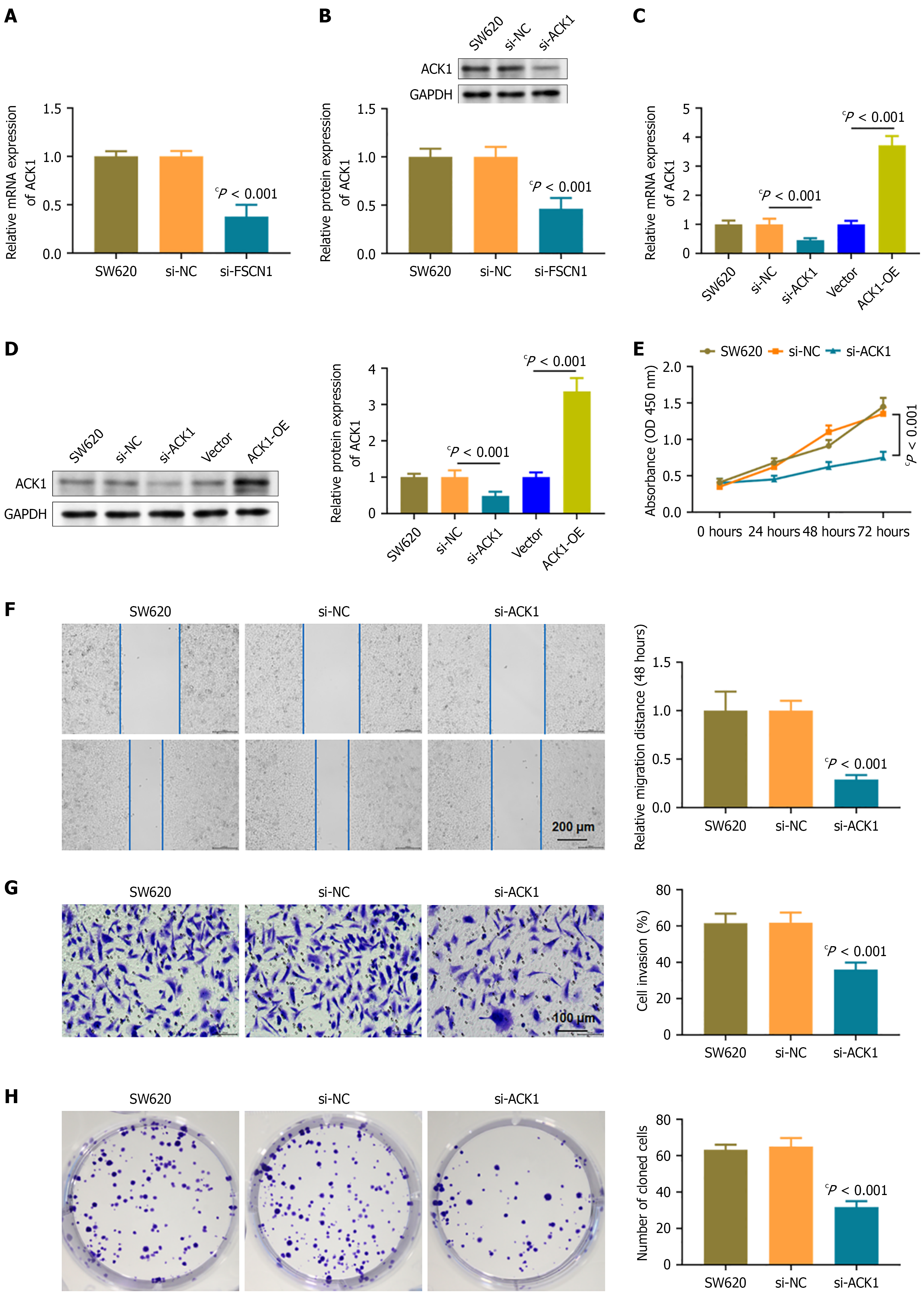
Figure 4 Fascin actin-bundling protein-1 positively regulates activated Cdc42-associated kinase 1 expression and activated Cdc42-associated kinase 1 promotes malignant phenotype of colon cancer cells.
A and B: The mRNA and protein expression levels of activated Cdc42-associated kinase 1 (ACK1) after fascin actin-bundling protein-1 knockdown; C and D: Detection of ACK1 overexpression and knockdown (si-ACK1) efficiency; E: Cell proliferation; F and G: Transwell cell migration and invasion. Scale bar = 200 μm and 100 μm; H: Clone formation experiment. cP < 0.001. ACK1: Activated Cdc42-associated kinase 1; FSCN1: Fascin actin-bundling protein-1; ACK1-OE: Activated Cdc42-associated kinase 1 overexpression; NC: Non-targeting control.
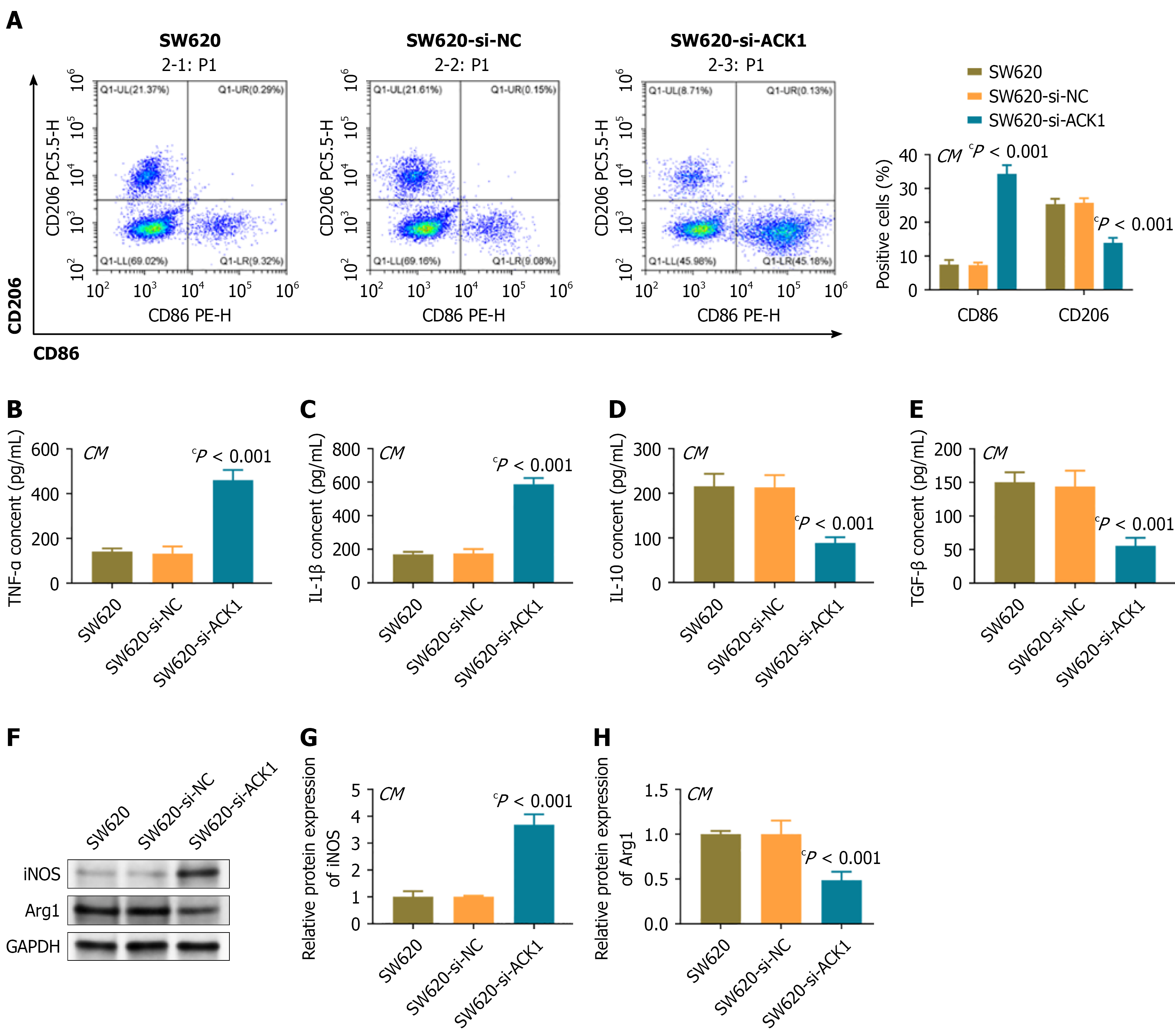
Figure 5 Activated Cdc42-associated kinase 1 silencing inhibits M2 phenotype of macrophages.
A: Flow cytometry analysis of the ratio of CD86+ (M1) and CD206+ (M2) in macrophages; B-E: Enzyme-linked immunosorbent assay was used to detect the secretion levels of M1 and M2 macrophage cytokines; F-H: Protein expression of inducible nitric oxide synthase and arginase 1. cP < 0.001. PMA induced differentiation of THP-1 macrophages was treated with conditioned medium containing SW620, SW620-si-NC, and SW620-si-CK1 cells, respectively. TNF-α: Tumor necrosis factor-α; TGF-β1: Transforming growth factor-β1; iNOS: Inducible nitric oxide synthase; Arg1: Arginase 1; ACK1: Activated Cdc42-associated kinase 1; NC: Non-targeting control; CM: Conditioned medium; IL: Interleukin.
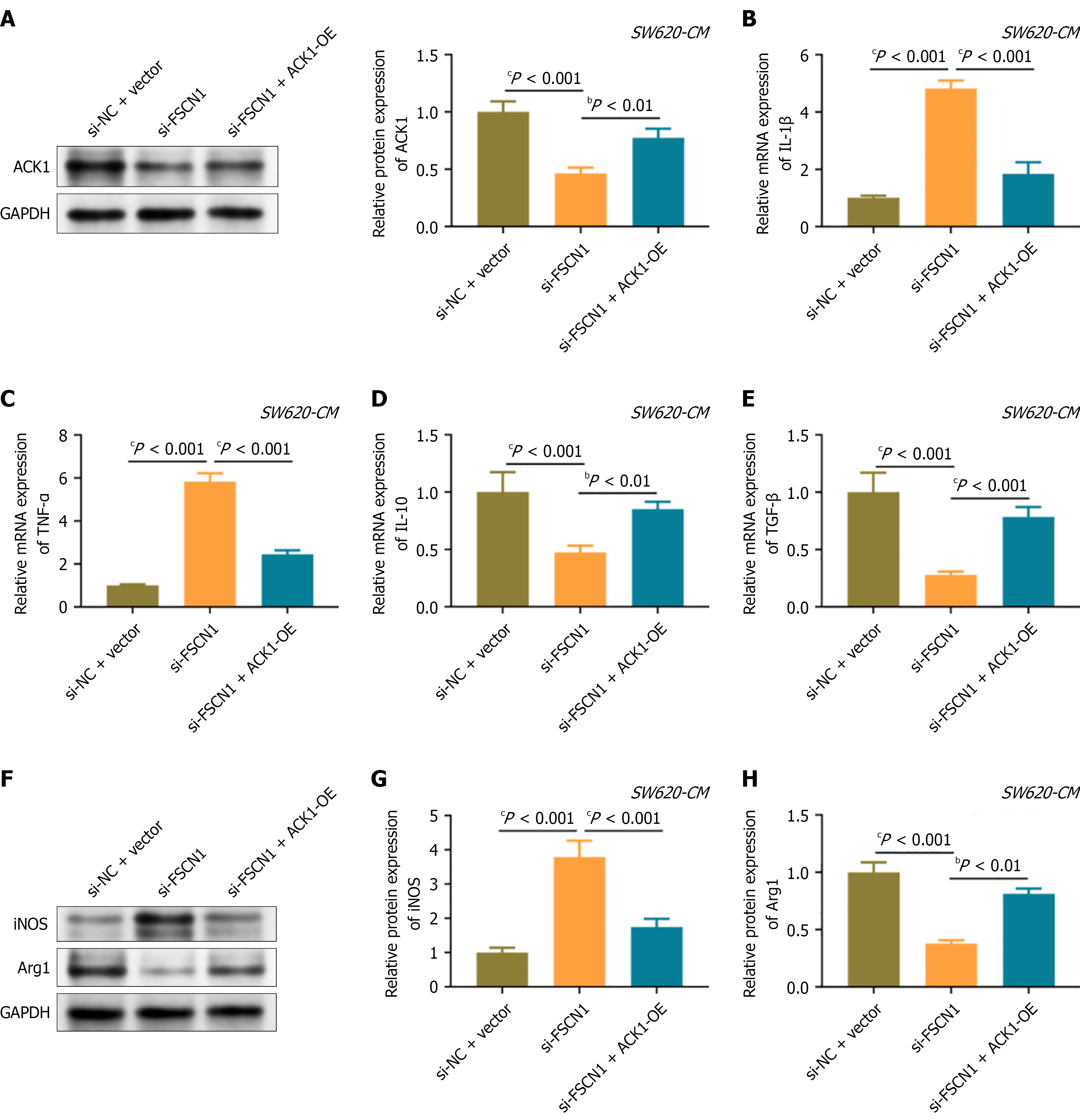
Figure 6 Fascin actin-bundling protein-1 regulates macrophage polarization through activated Cdc42-associated kinase 1.
A: Activated Cdc42-associated kinase 1 protein expression; B-E: The mRNA expression levels of M1 and M2 macrophage cytokines; F-H: Protein expression of Inducible nitric oxide synthase and arginase 1. bP < 0.01. cP < 0.001. FSCN1: Fascin actin-bundling protein-1; TNF-α: Tumor necrosis factor-α; TGF-β1: Transforming growth factor-β1; iNOS: Inducible nitric oxide synthase; Arg1: Arginase 1; ACK1: Activated Cdc42-associated kinase 1; NC: Non-targeting control; ACK1-OE: Activated Cdc42-associated kinase 1 overexpression; CM: Conditioned medium; IL: Interleukin.
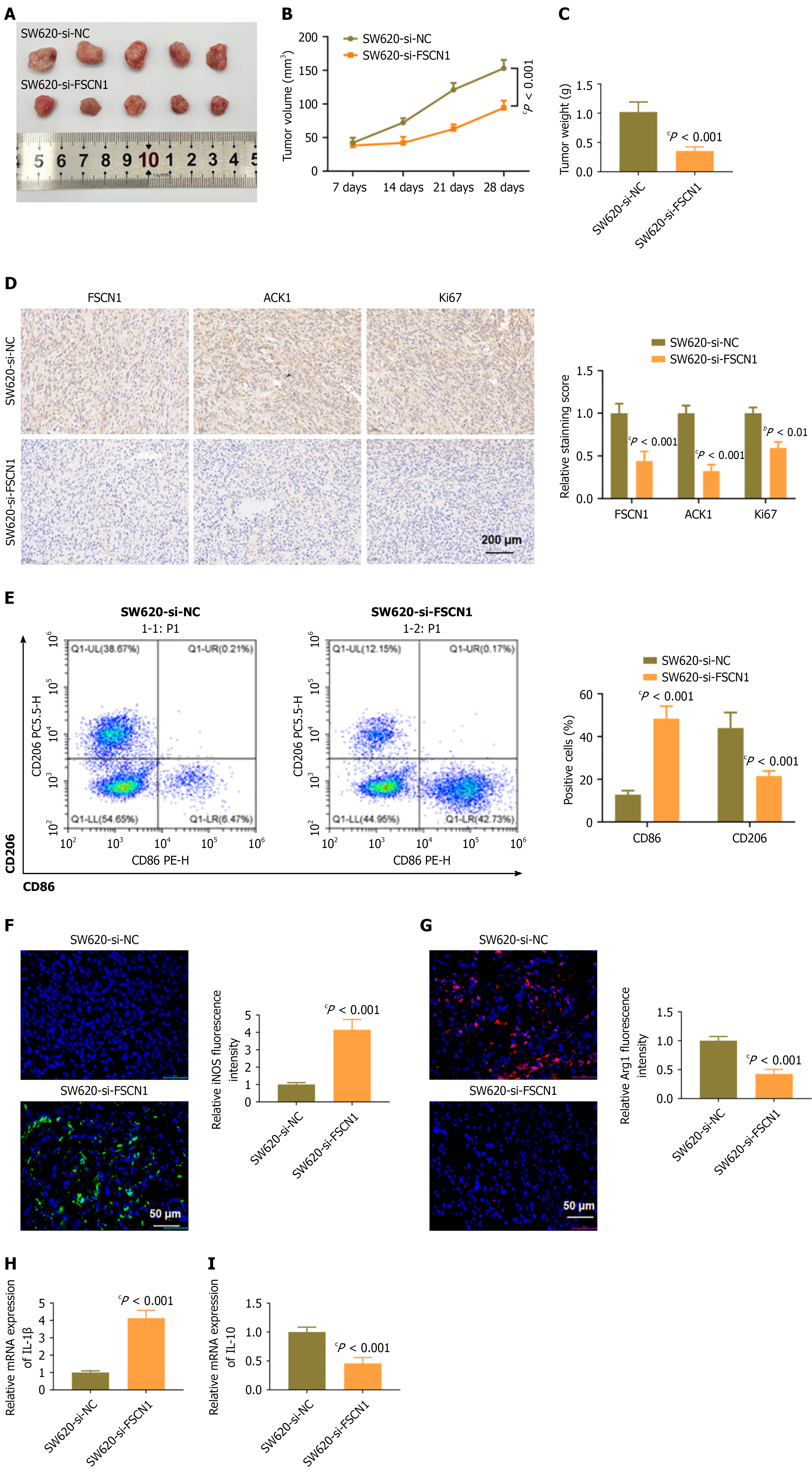
Figure 7 Fascin actin-bundling protein-1 silencing inhibits colon cancer growth and reshapes the tumor immune microenvironment in vivo.
A: Representative tumor photos; B: Tumor growth curve; C: End point tumor weight; D: Immunohistochemistry staining of fascin actin-bundling protein-1, activated Cdc42-associated kinase 1, and Ki67. Scale bar = 200 μm; E: Flow cytometry analysis of polarization status of macrophages in tumors; F and G: Immunofluorescence staining images of Inducible nitric oxide synthase and arginase 1, with DAPI counterstaining of the cell nucleus. Scale bar = 50 μm; H and I: The mRNA levels of interleukin-1β and interleukin-10. bP < 0.01. cP < 0.001. FSCN1: Fascin actin-bundling protein-1; iNOS: Inducible nitric oxide synthase; Arg1: Arginase 1; ACK1: Activated Cdc42-associated kinase 1; NC: Non-targeting control; IL: Interleukin.
- Citation: Wang Z, Liu C, Liu H, Jiang Y, Ma Y. FSCN1/ACK1 axis-dependent M2 macrophage polarization fuels colorectal cancer progression. World J Gastroenterol 2026; 32(44): 121025
- URL: https://www.wjgnet.com/1007-9327/full/v32/i44/121025.htm
- DOI: https://dx.doi.org/10.3748/wjg.121025