Published online May 26, 2019. doi: 10.12998/wjcc.v7.i10.1133
Peer-review started: November 16, 2018
First decision: January 12, 2019
Revised: April 23, 2019
Accepted: May 1, 2019
Article in press: May 2, 2019
Published online: May 26, 2019
Processing time: 192 Days and 0.4 Hours
Spasticity affects a large number of children, mainly in the setting of cerebral palsy, however, only a few paediatric neurosurgeons deal with this problem. This is mainly due to the fact that until 1979, when Fasano has published the first series of selective dorsal rhizotomy (SDR), neurosurgeons were able to provide such children only a modest help. The therapy of spasticity has made a great progress since then. Today, peroral drugs, intramuscular and intrathecal medicines are available, that may limit the effects of the disease. In addition, surgical treatment is gaining importance, appearing in the form of deep brain stimulation, peripheral nerve procedures and SDR. All these options offer the affected children good opportunities of improving the quality of life.
A 15-year old boy is presented that was surgically treated for spasticity as a result of cerebral palsy. Laminotomy at L1 level was performed and L1 to S1 nerve roots were isolated and divided in smaller fascicles. Then, the SDR was made.
We describe a patient report and surgical technique of SDR that was performed in Slovenia for the first time.
Core tip: Spasticity affects a large number of children, mainly in the setting of cerebral palsy. The therapy of spasticity has made a great progress. Today, peroral drugs, intramuscular and intrathecal medicines are available. In addition, surgical treatment is gaining importance. We describe a patient report and surgical technique of SDR that was performed in Slovenia for the first time and successfully introduced in the clinical practice.
- Citation: Velnar T, Spazzapan P, Rodi Z, Kos N, Bosnjak R. Selective dorsal rhizotomy in cerebral palsy spasticity - a newly established operative technique in Slovenia: A case report and review of literature. World J Clin Cases 2019; 7(10): 1133-1141
- URL: https://www.wjgnet.com/2307-8960/full/v7/i10/1133.htm
- DOI: https://dx.doi.org/10.12998/wjcc.v7.i10.1133
Spasticity is a motor disorder characterized by an increase in muscle tone that inter-feres with the mobility and therefore affects the quality of life[1]. Spasticity, which results from the unbalanced factors that act on rising and lowering the muscle tone, is the result of an abnormal increase and irritability of myotactic reflexes and depends on the speed of movement. Although spasticity is evident in various diseases that affect muscle tone, such as idiopathic or secondary forms, mainly in connection with dystonia or atethosis, it is most commonly associated with cerebral palsy, which affects 2 to 3 children in 1000 people[2]. The spasticity in cerebral palsy is not evident immediately at birth. On the contrary, it usually becomes pronounced only after the first year of life. The treatment of spasticity is important to reduce the muscle tone, improve the quality of life and reduce pain and deformations. It has two objectives: to reduce the efferent impulses that stimulate muscles and rise the abnormal muscle tone [with surgical procedures such as selective dorsal rhizotomy (SDR)] or increase the inhibitory activity on the muscle tone (with the action of drugs such as baclofen)[2-4].
Spasticity typically affects some muscle groups more than others[3]. In the clinical picture of a child with a cerebral palsy, muscular rigidity, tiredness, muscle pain and cramps are evident. The pain and muscle cramps are particularly pronounced in the nocturnal hours. In time, the spasticity may also lead to the deformations of the skeleton, such as luxations and subluxations of the hip and ankle joints. Additionally, upper limbs are almost always affected to some degree. The most affected are abductors and flexors. This is evident in patients with spastic paraparesis, who are standing on the toes with flexed hips and knees and internal rotation of lower limbs[4-6].
Clinically, spasticity is described according to the affected limbs as spastic tetraparesis, spastic diplegia (or paraparesis), spastic haemy- and monoparesis[2,4]. The most used evaluation scale for spasticity is Ashworth Scale (and very similar Modified Ashworth Scale), which determinates the degree of spasticity (Table 1). Spasticity can also affect paraspinal muscles, but this disorder is not included in the above classification. The motor function, which may also be severely affected due to the spasticity of the muscles, is classified according to the Gross Motor Function Classification System (GMFCS) (Table 2)[4-6].
| SCORE- Degree of spasticity | DESCRIPTION |
| 0 | No increase in muscle tone. |
| 1 | Slight increase in tone giving a catch when the limb was moved in flexion or extension. |
| 2 | More marked increase in tone but limb easily flexed. |
| 3 | Considerable increase in tone - passive movement difficult. |
| 4 | Limb rigid in flexion or extension. |
| SCORE- Level of spasticity | DESCRIPTION |
| Level I | Walks well in all settings. Balance and speed may be limited compared with children developing normally. |
| Level II | Walks in most settings but may have difficulty walking long distances or with balance. May utilise personal or environmental mobility aids to climb stairs. |
| Level III | Walks with the use of hand-held mobility aids such as K-walkers in most indoor settings. Uses wheeled mobility for longer distance travel. |
| Level IV | Utilises wheeled mobility aids in most settings (either attendant-propelled or powered) and requires assistance to transfer. |
| Level V | Transported in wheelchairs in all settings and has limited to no antigravity head, trunk and limb control. |
There are many forms of spasticity treatment. The aim is to reduce the degree of spasticity, rather than cure it completely[7]. The treatment encompasses many modalities that include oral medications, intramuscular injections of intrathecal medications, physiotherapy and orthopaedic procedures and even neurosurgical operations[8]. One of the options includes SDR. It is a neurosurgical procedure with the aim of reducing spasticity and improving mobility in children with cerebral palsy suffering from lower extremity spasticity[9-11]. The technique involves a selective division of the lumbosacral sensory rootlets with the aid of intraoperative neurophysiological monitoring. The SDR represents a valuable surgical option especially for young patients with bilateral spastic cerebral palsy with the objective of improving lower limb function[10].
A proper form of treatment should be therefore proposed by a multidisciplinary team with experience in each of these therapies that assesses the patients. The most important factor in predicting the SDR performance is not so much a sample of neurophysiological monitoring, acquired during the operation, but the correct preoperative selection of the patient[12]. Choosing the correct patient for SDR is therefore of utmost importance and requires a comprehensive evaluation. Ideal patients for SDR are children between 4 and 8 years of age, with spastic paraparesis, which impedes their pattern of walking. Such patients usually have preserved strength in the lower extremities and only rare contractions. Unfortunately, only few children with cerebral palsy fulfil these requirements, in clinical practice less than 5%[13-15].
Although SDR has been efficacious in treating spastic patients, many clinicians are not well aware of the procedure, its indications and expected outcomes due to the limited number of centres performing this procedure[10,16]. In this article, the authors describe the SDR technique in the management of spastic diplegia in a child with cerebral palsy that was performed in Slovenia for the first time and has been introduced in the neurosurgical practice.
A 15-year old boy was admitted to the neurosurgery department for SDR. He was experiencing severe and painful muscle cramps and the increased muscle tone prevented him from normal walking and sitting.
A 15-year old boy was admitted to the neurosurgery department for SDR. He was born prematurely at the gestational age of 35 wk and postnatally sustained an intraventricular haemorrhage, grade 2. Later on, he was recovering well. There were no signs of hydrocephalus and he was regularly followed-up at paediatric neurology and physiatry. He was cognitively intact. After one year, an increased muscle tone was observed and he was included into a rehabilitation programme. A spastic diplegia started to develop and the muscle tone has increased progressively. For many years, the physiatric, orthopaedic and medicamentous care sufficed to counterbalance the spasticity, however, due to deformations and shortage of hamstrings and calf muscles he needed an orthopaedic intervention. At the age of 15, he was referred to the neurosurgeon as a result of increasing spasticity and difficulties in walking, sitting and numerous muscle cramps in lower extremities.
No past illnesses were documented in the new-born or mother.
Unremarkable.
At the examination, the cranial nerves and upper extremities were functioning normally. In the lower extremities, however, spasticity was evident and the muscle tone was rated at 4, according to the Modified Ashworth Scale and to 3 according to the GMFCS. The spastic gait was evident as was the scissor-pattern walk. The sphincters were unaffected. After initial neurosurgical evaluation, a multidisciplinary team assessed and discussed possible treatment options. For muscle tone reduction, the SDR was recommended.
Laboratory examinations were within the normal range. The haemostasis was normal, as was the blood count and the biochemistry test.
No special imaging was required for the surgical treatment. The level L1 was marked with x-rays intraoperatively for the location of conus medullaris.
The final diagnosis was consistent with the spasticity as a result of cerebral palsy.
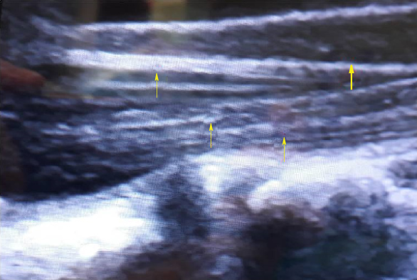
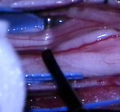
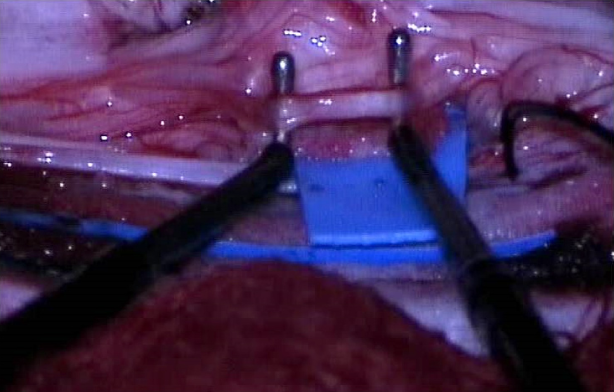
After initial anaesthesiological preparation, the boy was placed prone on the operating table. The neuromonitoring for dermatomes and myotomes L2 to S2 was attached. The level L1 was marked with X-rays for the location of conus medullaris. A laminotomy L1 followed and the dural sac was exposed. With the ultrasound, the location of the conus and cauda was determined and these structures were clearly seen (Figure 1). Under the operating microscope, the dura was cut in the median line and the arachnoid dissected. The conus was exposed, surrounded by the spinal roots and left and right roots were separated. On the left side, L1 root was identified anatomically, exiting from the neuroformanen. The root is composed of three fascicles, with the most ventral one being motor and the two caudal ones sensory. These two fascicles were then separated from the motor part and one of these two rootlets was coagulated and cut, so as to disconnect 50% of the fibres (Figure 2).
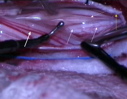
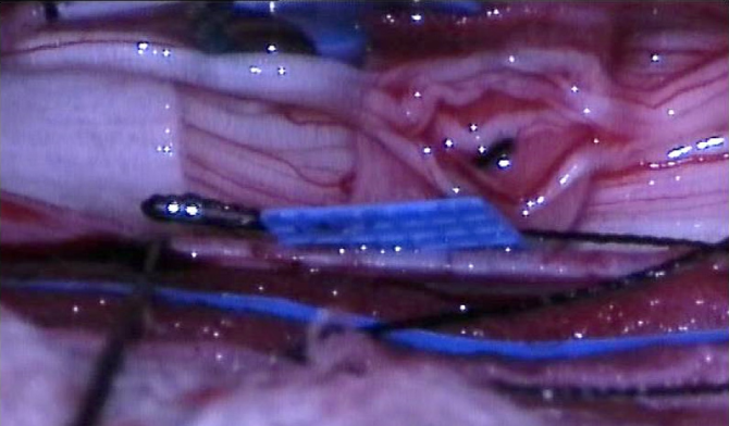
Disconnection of the lower roots then followed. First, the groove separating the motor and sensory roots was identified anatomically and electrophysiologically (Figure 3). The motor roots trigger the EMG potential when touched with an instrument, on the contrary to the sensory roots. Then, the sensory roots from L2 to S2 were separated from other structures with an elastic cloth (4cm × 1.5cm). The limit between S2 and S3 roots was anatomically recognized, since the S3 roots are smaller and thinner than the S2. The L2 root, which is located most laterally, was then isolated and confirmed with the electrophysiology. This root was again separated from others with a fine cotton patty and divided into four fascicles with a fine blunt microdiscector (Figure 4). Neurophysiologically the most abnormally active fascicles were coagulated and disconnected (Figure 5). The root L2 was then placed separately from other lower lumbar and sacral roots. The same procedure was exerted on roots L3 to S1. The number of fascicles was various, ranging from four to seven, and two-thirds of them were cut, specifically those with an abnormal electrophysiological response. The S2 root required a special attention. It was divided in two fascicles and the potential of the pudendal action potential was explored. Here, part of the fascicle with lower electrophysiological sphincter activity was cut. An identical procedure was repeated on the right side.
A meticulous intradural haemostasis followed and the dura was closed with a running suture. The lamina was positioned and attached on the place with sutures and the wound was closed in layers.
After the operation, the patient was transferred to the neurosurgical intensive care and the next day onto the ward. The muscle tone was reduced in a day after surgery, as was the strength in the lower extremities. It was not reduced to such extent, however to prevent the patient from walking. No motor deficits were observed and the sphincter control was intact. The pain subsided gradually in as well as the muscle cramps in the lower extremities. The physiotherapy started gradually after the established protocol from the first day to the discharge, first with passive and then with active exercises, gait control and walking. A special emphasis was put into verticalisation, initially sitting on the wheelchair and then progressive training of proper walking. The patient did not experienced difficulties during this part of rehabilitation.
Ten days after the operation he was discharged to specialised rehabilitation institute for further treatment. The Ashworth Scale and the GMFCS at the discharge were both rated at 2. The neurosurgical examination was planned after on month and the general condition improved markedly. The boy was able to walk independently, the muscle tone was lower (Modified Ashowrth Scale and the GMFCS were 2). No sensory and sphincter disturbances were reported. Next neurosurgical evaluation has been planned after three and six months, as well as continuous physiatric assessment.
Spasticity in cerebral palsy is still a great challenge for patients and clinicians treating them, despite the advances in medical care, rehabilitation medicine and surgical techniques. The dorsal rhizotomy as an option for spasticity treatment is not new, as it has been practiced for over 100 years[3-5,17]. First described in 1908 by Sherrington and later developed and performed also by Foerster, early surgical procedures were effective in reducing spasticity. Despite favourable results on muscle tone, they were associated with significant postoperative morbidity as a result of sensory loss and ataxia, which occurred in the total section of lumbar sensory roots (usually section encompassed the L2 to S1 roots). This has led to a temporary omission of the technique for several decades, until in 1979, Fasano described the selective section of the lumbar sensory roots, known as SDR. Here, only those fascicles were disconnected that were causing a permanent and pathological contraction of the muscle during the neurophysiological stimulation[7]. Later, Peacock defined the significance of the pathological response to the electrophysiological irritation of the dorsal fascicle even more precisely. Since then, various modifications of the original surgical technique have been described. Currently, SDR is one of most commonly performed ablative treatment procedures for spasticity in cerebral palsy in children. Technical advancements over the last two decades have helped to reduce the invasiveness of the procedure even more[17-19].
In Slovenia, the SDR has been performed for the first time in October 2017 and since then it was introduced in the regular paediatric neurosurgical activity. Until then, the children with spasticity due to the cerebral palsy were referred for surgical treatment to other centres abroad that performed this operation, most commonly to St. Louis, Missouri, United States. The initial evaluation and medical treatment were done in the Slovenian rehabilitation institute, as well as the postoperative and late rehabilitation, eventual orthopaedic corrections and continuous physiotherapy. The neurosurgical technique that was used in our patient was the same as already described in detail by Sindou et al[20]. The laminotomy at L1 level offers an unob-structed view to the end part of the conus and the cauda and the identification of the sensory roots that need to be divided, monitored and cut, namely the roots form L1 to S2 bilaterally. The SDR can also be performed through a larger laminotomy, from L1 to S1. Using this approach each dorsal root can be recognized at the correspondent neuroforamen. This wide laminotomy offers a good view but. However, it may compromise the stability of the spine and with time lead to scoliosis[20,21]. In our case, the bone fragment was sutured in place at the end of the operation to prevent possible instability.
Monitoring is of vital importance and helps the surgeons to confirm the radix that is going to be cut[22]. Normally, each sensory root is divided into 4 to 8 rootlets, each of them is monitored and those evoking the most abnormal response in the muscles are cut, which is usually about 70% of the rootlets. It is particularly important to spare the innervation of the sphincters and care must be taken during the operation not to damage these nerves. The pudendal action potential is of great usefulness for recognition of these rootlets[19-24].
The spasticity in cerebral palsy is the most common indication for SDR[10,19-25]. This diagnosis may be set when a child at a certain age does not achieve a normal motor development (e.g., head lifting, sitting, standing and walking). The gait and posture are inappropriate with an increased muscle tone. In the initial phase, the muscles are loose, with spasticity occurring later. In the early childhood, the typical signs of cerebral palsy may not be evident and the spasticity may occur later, usually after 6 mo of age. In general, the symptoms may be diverse, ranging from almost undetectable clumsiness to severe disability. Typical symptoms, regardless of type, include abnormal muscle tone, balance disorders and abnormally lively tendon reflexes, often accompanied by involuntary movements. The walk is unstable, usually scissor pattern of walking may be observed or walking on toes. As a result of increased muscle tone, the deformation of bones and joints may also occur, worsening the mobility[10,13]. Cerebral palsy can also be accompanied with epileptic seizures, apraxia, sensory disorders and disturbances in sphincter control. Pain, which occurs in combination with a spastically elevated muscular tone, abnormal posture and hardened joints, can often be associated with these symptoms. In the spastic form of the cerebral palsy, which is the most common, the muscular tone is extremely elevated and the tendon reflexes are very pronounced. Muscle contractions may sometimes be provoked by an unexpected hearing or visual stimulus. Joint contractions worsen the general condition and improve to deformations and pain. Swallowing and speech disorders are not uncommon in this type of cerebral palsy[18-24].
According to the affected limbs and spasticity, the cerebral palsy may be divided into a paraplegic, diplegic and tetraplegic type. The paraplegic form is most often the result of a reduced blood supply to the child's brain in the third trimester of pregnancy. Rarely diagnosed at birth, it usually manifests at the age of 6 months. It is this type that is most suitable for the SDR, as was also the case in our patient[7,26].
The management of cerebral palsy is complex and therefore requires a multidisciplinary approach, which should be tailored for individual cases. As the SDR is one of ablative surgical therapies with irreversible properties, preoperative assessment and decision making for the operation are critical[22,27]. Not all patients with cerebral palsy suffering from spasticity need treatment. The highest success has been observed in patients with Modified Ashworth grades 2 and 3. In low degrees of spasticity, the daily activities are not disturbed to such degree, that the operation would bring a relief, as it is with high grades. The goals of the treatment include improvement of child’s functional state, especially the pattern of walking; facilitate the everyday care, to prevent the appearance of contractures and deformations and the onset of pain. The SDR is mostly suitable for children with spastic diplegia who are potential ambulators[27,28]. When choosing the best possible treatment, the main considerations include the age of the child and the goals for the improvement of the general condition. When considering the age, the SDR is ideal procedure from 4 to 8 years, as the growth and brain myelinization is almost complete during this time. Rarely, the indication for SDR is justified after 15 years of age, since it is difficult to strengthen the muscles and alter the pattern of walking after the surgery. Though, recent articles have showed a positive effect on spasticity also in more severely affected patients (Modified Ashworth Scale 4 and 5)[29,30]. After the SDR, persistent deformations and tendons should be corrected by appropriate orthopaedic surgery. The motivation of the child and parents is a fact that always must to be considered before the surgery[26-29].
Because of pronounced spasticity and ambulatory inability, we have decided to treat the spastic diplegia in our patient, despite his age. However, after the SDR, the treatment of patients is not completed[5,6]. The spasticity immediately after the operation declines and the muscle strength deteriorates, thus worsening the condition transiently. This usually lasts for approximately three weeks[13]. During this time, paraesthesia and burning or painful sensations tin extremities can be noted. Gradually, they disappear, the muscle tone clearly lowers and the pattern of walking improves. In our patient, the spasticity was reduced immediately and dropped from Modified Ashworth grade 4 to 2. There was neither paraesthesia or pain reported nor sphincter worsening.
In the post-operative phase, the rehabilitation plays an essential role, supplemented by possible corrective orthopaedic interventions. Due to the inhibitory effect on ascendant interneurons, also the spasticity of the upper limbs can be reduced and the effect on bladder control can be improved, as well as the general functioning[13,24].
The operation in our patient was successful and the recovery was smooth. In the future, more patents with spastic diplegia will be included into the multidisciplinary programme for spasticity treatment, employing the SDR technique, thus improving the life of cerebral paralysis patients.
The SDR is an efficient surgical technique for reducing spasticity, especially in young children with spastic diplegia as a result of cerebral palsy. The technique requires meticulous neuromonitoring during the operation and continuous child rehabilitation with long-term follow-up. The treatment outcome of the SDR is long-lasting and the side effects with careful surgery are minimal.
Manuscript source: Invited manuscript
Specialty type: Medicine, Research and Experimental
Country of origin: Slovenia
Peer-review report classification
Grade A (Excellent): 0
Grade B (Very good): B
Grade C (Good): C, C
Grade D (Fair): 0
Grade E (Poor): 0
P-Reviewer: Chowdhury FH, Vaudo G, Yang XJ S-Editor: Dou Y L-Editor: A E-Editor: Xing YX
| 1. | Lieber RL, Roberts TJ, Blemker SS, Lee SSM, Herzog W. Skeletal muscle mechanics, energetics and plasticity. J Neuroeng Rehabil. 2017;14:108. [RCA] [PubMed] [DOI] [Full Text] [Full Text (PDF)] [Cited by in Crossref: 76] [Cited by in RCA: 98] [Article Influence: 12.3] [Reference Citation Analysis (0)] |
| 2. | O'Shea TM. Diagnosis, treatment, and prevention of cerebral palsy. Clin Obstet Gynecol. 2008;51:816-828. [RCA] [PubMed] [DOI] [Full Text] [Full Text (PDF)] [Cited by in Crossref: 146] [Cited by in RCA: 106] [Article Influence: 6.2] [Reference Citation Analysis (0)] |
| 3. | Nahm NJ, Graham HK, Gormley ME, Georgiadis AG. Management of hypertonia in cerebral palsy. Curr Opin Pediatr. 2018;30:57-64. [RCA] [PubMed] [DOI] [Full Text] [Cited by in Crossref: 29] [Cited by in RCA: 42] [Article Influence: 6.0] [Reference Citation Analysis (1)] |
| 4. | D'Aquino D, Moussa AA, Ammar A, Ingale H, Vloeberghs M. Selective dorsal rhizotomy for the treatment of severe spastic cerebral palsy: efficacy and therapeutic durability in GMFCS grade IV and V children. Acta Neurochir (Wien). 2018;160:811-821. [RCA] [PubMed] [DOI] [Full Text] [Cited by in Crossref: 26] [Cited by in RCA: 35] [Article Influence: 5.0] [Reference Citation Analysis (0)] |
| 5. | Roberts A, Stewart C, Freeman R. Gait analysis to guide a selective dorsal rhizotomy program. Gait Posture. 2015;42:16-22. [RCA] [PubMed] [DOI] [Full Text] [Cited by in Crossref: 23] [Cited by in RCA: 13] [Article Influence: 1.3] [Reference Citation Analysis (0)] |
| 6. | Roberts A. Surgical management of spasticity. J Child Orthop. 2013;7:389-394. [RCA] [PubMed] [DOI] [Full Text] [Cited by in Crossref: 20] [Cited by in RCA: 18] [Article Influence: 1.5] [Reference Citation Analysis (0)] |
| 7. | Grunt S, Fieggen AG, Vermeulen RJ, Becher JG, Langerak NG. Selection criteria for selective dorsal rhizotomy in children with spastic cerebral palsy: a systematic review of the literature. Dev Med Child Neurol. 2014;56:302-312. [RCA] [PubMed] [DOI] [Full Text] [Cited by in Crossref: 53] [Cited by in RCA: 62] [Article Influence: 5.6] [Reference Citation Analysis (0)] |
| 8. | Graham D, Aquilina K, Mankad K, Wimalasundera N. Selective dorsal rhizotomy: current state of practice and the role of imaging. Quant Imaging Med Surg. 2018;8:209-218. [RCA] [PubMed] [DOI] [Full Text] [Cited by in Crossref: 12] [Cited by in RCA: 15] [Article Influence: 2.1] [Reference Citation Analysis (0)] |
| 9. | Sitthinamsuwan B, Nunta-Aree S. Ablative neurosurgery for movement disorders related to cerebral palsy. J Neurosurg Sci. 2015;59:393-404. [PubMed] |
| 10. | Farmer JP, Sabbagh AJ. Selective dorsal rhizotomies in the treatment of spasticity related to cerebral palsy. Childs Nerv Syst. 2007;23:991-1002. [RCA] [PubMed] [DOI] [Full Text] [Cited by in Crossref: 51] [Cited by in RCA: 49] [Article Influence: 2.7] [Reference Citation Analysis (0)] |
| 11. | Engsberg JR, Park TS. Selective dorsal rhizotomy. J Neurosurg Pediatr. 2008;1:177; discussion 178-177; discussion 179. [RCA] [PubMed] [DOI] [Full Text] [Cited by in Crossref: 2] [Cited by in RCA: 2] [Article Influence: 0.1] [Reference Citation Analysis (0)] |
| 12. | Oki A, Oberg W, Siebert B, Plante D, Walker ML, Gooch JL. Selective dorsal rhizotomy in children with spastic hemiparesis. J Neurosurg Pediatr. 2010;6:353-358. [RCA] [PubMed] [DOI] [Full Text] [Cited by in Crossref: 22] [Cited by in RCA: 23] [Article Influence: 1.5] [Reference Citation Analysis (0)] |
| 13. | Langerak NG, Lamberts RP, Fieggen AG, Peter JC, Peacock WJ, Vaughan CL. Functional status of patients with cerebral palsy according to the International Classification of Functioning, Disability and Health model: a 20-year follow-up study after selective dorsal rhizotomy. Arch Phys Med Rehabil. 2009;90:994-1003. [RCA] [PubMed] [DOI] [Full Text] [Cited by in Crossref: 37] [Cited by in RCA: 40] [Article Influence: 2.5] [Reference Citation Analysis (0)] |
| 14. | Park TS, Liu JL, Edwards C, Walter DM, Dobbs MB. Functional Outcomes of Childhood Selective Dorsal Rhizotomy 20 to 28 Years Later. Cureus. 2017;9:e1256. [RCA] [PubMed] [DOI] [Full Text] [Full Text (PDF)] [Cited by in Crossref: 13] [Cited by in RCA: 23] [Article Influence: 2.9] [Reference Citation Analysis (0)] |
| 15. | Park TS, Edwards C, Liu JL, Walter DM, Dobbs MB. Beneficial Effects of Childhood Selective Dorsal Rhizotomy in Adulthood. Cureus. 2017;9:e1077. [RCA] [PubMed] [DOI] [Full Text] [Full Text (PDF)] [Cited by in Crossref: 6] [Cited by in RCA: 20] [Article Influence: 2.5] [Reference Citation Analysis (0)] |
| 16. | Park TS, Johnston JM. Surgical techniques of selective dorsal rhizotomy for spastic cerebral palsy. Technical note. Neurosurg Focus. 2006;21:e7. [PubMed] |
| 17. | Aquilina K, Graham D, Wimalasundera N. Selective dorsal rhizotomy: an old treatment re-emerging. Arch Dis Child. 2015;100:798-802. [RCA] [PubMed] [DOI] [Full Text] [Cited by in Crossref: 39] [Cited by in RCA: 52] [Article Influence: 5.2] [Reference Citation Analysis (0)] |
| 18. | Narayanan UG. Management of children with ambulatory cerebral palsy: an evidence-based review. J Pediatr Orthop. 2012;32 Suppl 2:S172-S181. [RCA] [PubMed] [DOI] [Full Text] [Cited by in Crossref: 68] [Cited by in RCA: 69] [Article Influence: 5.3] [Reference Citation Analysis (0)] |
| 19. | Lynn AK, Turner M, Chambers HG. Surgical management of spasticity in persons with cerebral palsy. PM R. 2009;1:834-838. [RCA] [PubMed] [DOI] [Full Text] [Cited by in Crossref: 30] [Cited by in RCA: 31] [Article Influence: 1.9] [Reference Citation Analysis (0)] |
| 20. | Sindou MP, Simon F, Mertens P, Decq P. Selective peripheral neurotomy (SPN) for spasticity in childhood. Childs Nerv Syst. 2007;23:957-970. [RCA] [PubMed] [DOI] [Full Text] [Cited by in Crossref: 63] [Cited by in RCA: 52] [Article Influence: 2.9] [Reference Citation Analysis (0)] |
| 21. | Steinbok P, Hicdonmez T, Sawatzky B, Beauchamp R, Wickenheiser D. Spinal deformities after selective dorsal rhizotomy for spastic cerebral palsy. J Neurosurg. 2005;102:363-373. [RCA] [PubMed] [DOI] [Full Text] [Cited by in Crossref: 10] [Cited by in RCA: 24] [Article Influence: 1.2] [Reference Citation Analysis (0)] |
| 22. | Tilton A. Management of spasticity in children with cerebral palsy. Semin Pediatr Neurol. 2009;16:82-89. [RCA] [PubMed] [DOI] [Full Text] [Cited by in Crossref: 38] [Cited by in RCA: 30] [Article Influence: 1.9] [Reference Citation Analysis (0)] |
| 23. | Steinbok P. Selective dorsal rhizotomy for spastic cerebral palsy: a review. Childs Nerv Syst. 2007;23:981-990. [RCA] [PubMed] [DOI] [Full Text] [Cited by in Crossref: 89] [Cited by in RCA: 79] [Article Influence: 4.4] [Reference Citation Analysis (0)] |
| 24. | Kothbauer KF, Deletis V. Intraoperative neurophysiology of the conus medullaris and cauda equina. Childs Nerv Syst. 2010;26:247-253. [RCA] [PubMed] [DOI] [Full Text] [Cited by in Crossref: 51] [Cited by in RCA: 57] [Article Influence: 3.8] [Reference Citation Analysis (0)] |
| 25. | Gump WC, Mutchnick IS, Moriarty TM. Selective dorsal rhizotomy for spasticity not associated with cerebral palsy: reconsideration of surgical inclusion criteria. Neurosurg Focus. 2013;35:E6. [RCA] [PubMed] [DOI] [Full Text] [Cited by in Crossref: 24] [Cited by in RCA: 30] [Article Influence: 2.7] [Reference Citation Analysis (0)] |
| 26. | Mittal S, Farmer JP, Al-Atassi B, Gibis J, Kennedy E, Galli C, Courchesnes G, Poulin C, Cantin MA, Benaroch TE. Long-term functional outcome after selective posterior rhizotomy. J Neurosurg. 2002;97:315-325. [RCA] [PubMed] [DOI] [Full Text] [Cited by in Crossref: 62] [Cited by in RCA: 54] [Article Influence: 2.3] [Reference Citation Analysis (0)] |
| 27. | Abou Al-Shaar H, Imtiaz MT, Alhalabi H, Alsubaie SM, Sabbagh AJ. Selective dorsal rhizotomy: A multidisciplinary approach to treating spastic diplegia. Asian J Neurosurg. 2017;12:454-465. [RCA] [PubMed] [DOI] [Full Text] [Full Text (PDF)] [Cited by in Crossref: 10] [Cited by in RCA: 12] [Article Influence: 1.5] [Reference Citation Analysis (0)] |
| 28. | Dudley RW, Parolin M, Gagnon B, Saluja R, Yap R, Montpetit K, Ruck J, Poulin C, Cantin MA, Benaroch TE, Farmer JP. Long-term functional benefits of selective dorsal rhizotomy for spastic cerebral palsy. J Neurosurg Pediatr. 2013;12:142-150. [RCA] [PubMed] [DOI] [Full Text] [Cited by in Crossref: 86] [Cited by in RCA: 88] [Article Influence: 7.3] [Reference Citation Analysis (0)] |
| 29. | Nemer McCoy R, Blasco PA, Russman BS, O'Malley JP. Validation of a care and comfort hypertonicity questionnaire. Dev Med Child Neurol. 2006;48:181-187. [RCA] [PubMed] [DOI] [Full Text] [Cited by in Crossref: 35] [Cited by in RCA: 34] [Article Influence: 1.8] [Reference Citation Analysis (0)] |
| 30. | Ingale H, Ughratdar I, Muquit S, Moussa AA, Vloeberghs MH. Selective dorsal rhizotomy as an alternative to intrathecal baclofen pump replacement in GMFCS grades 4 and 5 children. Childs Nerv Syst. 2016;32:321-325. [RCA] [PubMed] [DOI] [Full Text] [Cited by in Crossref: 28] [Cited by in RCA: 35] [Article Influence: 3.9] [Reference Citation Analysis (0)] |


