Copyright
©The Author(s) 2019.
World J Clin Cases. May 26, 2019; 7(10): 1133-1141
Published online May 26, 2019. doi: 10.12998/wjcc.v7.i10.1133
Published online May 26, 2019. doi: 10.12998/wjcc.v7.i10.1133
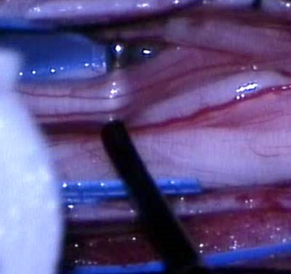
Figure 2 The exposure and isolation of the L1 root.
The root is composed of three fascicles, which will be subsequently separated. The motor fascicle will be spared, as will be one of the two sensory fascicles. The electrophysiological probe can be seen (right) and the cotton patty (left), which is used for isolation of the L1 root from other nerves.
- Citation: Velnar T, Spazzapan P, Rodi Z, Kos N, Bosnjak R. Selective dorsal rhizotomy in cerebral palsy spasticity - a newly established operative technique in Slovenia: A case report and review of literature. World J Clin Cases 2019; 7(10): 1133-1141
- URL: https://www.wjgnet.com/2307-8960/full/v7/i10/1133.htm
- DOI: https://dx.doi.org/10.12998/wjcc.v7.i10.1133


