Published online May 16, 2024. doi: 10.12998/wjcc.v12.i14.2426
Revised: February 22, 2024
Accepted: April 1, 2024
Published online: May 16, 2024
Processing time: 102 Days and 4.4 Hours
Various surgical techniques have been developed to enhance the nose shapes of Asian patients. Silicone implant augmentation rhinoplasty is widely used because it is relatively easy to perform and often yields satisfactory outcomes. However, this technique may lead to complications, including ischemia, necrosis, and over-augmentation. The most appropriate management of these complications, including infection, is immediate implant removal and revision surgery once the accompanying inflammation has healed. Occasionally, the patient may experience distress from nasal deformities during the intervention period.
Herein, we describe the case of a patient who underwent a secondary dorsal augmentation, with a folded dermofat graft harvested from the inguinal area and simultaneous implant removal, successfully preventing dimpling of the nasal deformity.
This surgical method can effectively manage implant-related complications following augmentation rhinoplasty using a silicone implant and provide satis
Core Tip: This case report presents noteworthy findings, as we introduce an innovative secondary rhinoplasty technique using a folded dermofat graft to address complications arising from silicone implant augmentation rhinoplasty. This procedure successfully maintained the nasal contour without complications after silicone implant augmentation rhinoplasty. This approach enhances patient satisfaction and provides a valuable solution to challenges associated with traditional methods, making it a promising option in managing complications following augmentation rhinoplasty.
- Citation: Kim H, Kim JH, Koh IC, Lim SY. Immediate secondary rhinoplasty using a folded dermofat graft for resolving complications related to silicone implants: A case report. World J Clin Cases 2024; 12(14): 2426-2430
- URL: https://www.wjgnet.com/2307-8960/full/v12/i14/2426.htm
- DOI: https://dx.doi.org/10.12998/wjcc.v12.i14.2426
Various surgical techniques have been developed in Asia to reshape flat, blunt noses into more desired shapes[1,2]. Among these, implant augmentation rhinoplasty has been widely used because it is simple to perform and can produce a wide range of alternative nose shapes[3]. However, this technique can also cause severe complications that require implant removal[4,5]. Correcting a nasal dimpling deformity that occurs following implant removal is challenging when the second procedure is performed following delays owing to complications. Herein, we present an immediate secondary rhinoplasty technique that minimizes dimpling in nasal deformities using a folded dermofat graft harvested from the inguinal area.
The patient complained of pain and discomfort in the nose.
Forty months after the surgery, the dorsum of the nose became erythematous and slightly inflamed, likely caused by excess pressure from the implant.
A 34-year-old woman without any underlying diseases underwent closed reduction of the nasal bone for a nasal bone fracture and simultaneous augmentation rhinoplasty with a silicone implant.
The patient had no significant past medical history or family history.
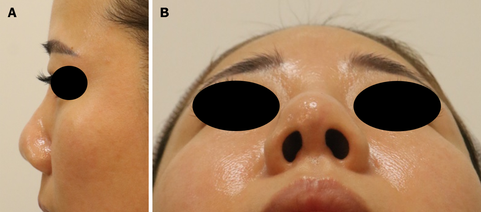
Upon admission, the contour of the implant became more conspicuous as the skin at the tip of the nose became thinner (Figure 1).
All results, including white blood cell count, erythrocyte sedimentation rate, and C-reactive protein level, were within the normal range.
Not applicable.
Nasal tip retraction with inflammation after augmentation rhinoplasty using a silicone implant.
We highly suspected an infection and a contracture and decided to remove the implant. We scheduled an immediate secondary rhinoplasty using a folded dermofat graft harvested from the inguinal area to replace the original implant.
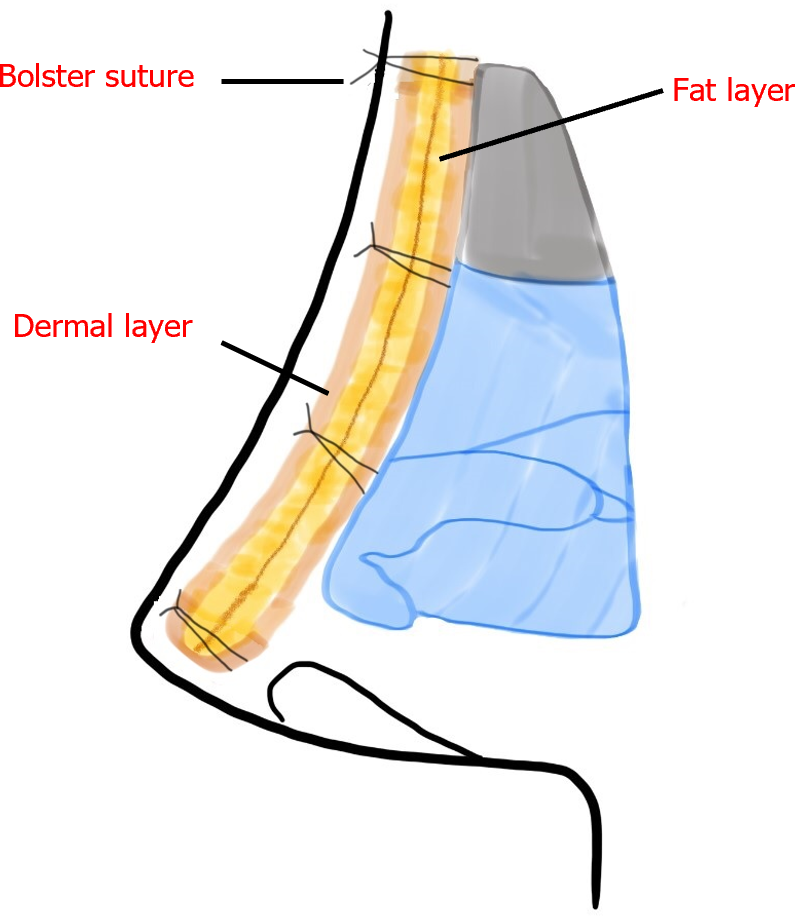
The distance from the nasal root to the nasal tip was 4.7 cm, and a graft double this length was designed for the folded dermofat graft. Using a transcolumellar approach, the implant was exposed by dissecting the cartilage and bone below the cartilaginous plane. The implant and adherent scar tissue were fully removed. The dermofat was harvested from the left inguinal area (approximately 1.0 cm × 9.5 cm) and folded using a 3-0 polydioxanone suture. The folded graft was inserted into the pocket of the nasal dorsum and fixed transcutaneously to the nasal root using a bolster suture (Figure 2). To prevent postoperative infection, a second-generation cephalosporin antibiotic was administered intravenously during the hospital stay for 2 d, followed by a 1-wk course of an oral first-generation cephalosporin antibiotic upon discharge. The columella and bolster stitches were removed on the 5th postoperative day, and those of the intranasal and donor sites were removed on the 12th postoperative day. The wound healed without any complications. Seven months later, the patient presented to our outpatient clinic. The shape of the nose was satisfactory, and no complications were noted (Figure 3).
Seven months later, the shape of the nose was satisfactory without any complications.
The nose of an Asian person is characterized by a limited projection of the nasal tip and an ala that is wider than the height of the nose[6]. Rhinoplasty is frequently performed on this nose type. Augmentation rhinoplasty is performed to increase the length and height of the dorsal and tip projections of the nose[2]. Augmentation rhinoplasty with silicone implants is the most widely performed procedure of this type because it helps achieve aesthetic goals for nose shapes relatively easily[3]. However, this surgery can sometimes result in complications, such as implant migration, contour irregularity, implant deviation, infection, extrusion, contracture, and skin lesions[4,5]. In particular, thinning of the skin due to foreign body reactions, scarring, and encapsulation around the implant may result in skin contracture, visibility of the contour of the implant, and even extrusion in severe cases[7].
In cases of complications accompanied by infection, the implant must be removed. However, determining the time for revision rhinoplasty can be challenging. A secondary rhinoplasty is recommended several months after implant removal after signs of infection are absent[8]. However, correction can be complex if dimpled nasal deformities, including excessive collapse and contracture, occur following implant removal. In the interim period, patients must tolerate this deformity and may experience psychological discomfort before undergoing a secondary rhinoplasty. Therefore, removing the implant and performing a secondary rhinoplasty concurrently is ideal.
Dermofat grafts have several advantages. For example, the autologous augmentation material comprises a full-thickness dermis and subcutaneous tissues. Since dermofat grafts are autologous tissues, they are relatively more susceptible to infection and foreign body reactions than other implant materials. The degree of volume maintenance at the surgical site may decrease over time. Depending on the location of the donor site, differences may occur between the thickness and fat density of the dermis[9,10]. In this study, we addressed these shortcomings using the “folded” dermofat method, which involves folding the collected dermofat in half. In general, the gluteal fold is the preferred donor site because of the presence of dense fat and a thick dermis[10,11]. However, the operative time may be prolonged when using this approach because the patient must be repositioned during the surgery. In addition to a longer surgical time, the cost burden to the patient is increased owing to the need for a longer duration of general anesthesia, and the patient may experience additional discomfort. The resultant scars can also be conspicuous if the patient wears a bikini. Because this part of the body continuously bears weight while sitting, wound healing can be slow and the incidence of complications, such as wound dehiscence, is high. When a dermofat graft is performed using the gluteal fold as the donor site, patients often complain of a blunt protruding nose because the dermis is excessively thick. For this procedure, we chose the inguinal area. Relatively large grafts can be obtained from this region, owing to the laxity of the skin, which makes it easier to create a more natural shape. Harvesting and grafting of dermofat can also be performed concurrently with the patient in the supine position, resulting in a very short operative time and high patient satisfaction. Because the dermis of the inguinal area is relatively thin[9], we were able to secure a satisfactory outcome with a sufficient nose height using this approach.
The folded dermofat graft method can be used to effectively manage implant complications following augmentation rhinoplasty with a silicone implant. In our case, a high level of patient satisfaction was achieved using this method.
Provenance and peer review: Unsolicited article; Externally peer reviewed.
Peer-review model: Single blind
Specialty type: Surgery
Country/Territory of origin: South Korea
Peer-review report’s scientific quality classification
Grade A (Excellent): 0
Grade B (Very good): B
Grade C (Good): 0
Grade D (Fair): 0
Grade E (Poor): 0
P-Reviewer: Xing HC, China S-Editor: Liu JH L-Editor: A P-Editor: Chen YX
| 1. | Wang X, Yan Q, Qiao Z, Deng Y, Li C, Sun Y, Xiong X, Meng X, Li W, Yi Z, Fang B. A New Definition for Alar Flare Based on "Alar Flare Angle". Aesthetic Plast Surg. 2023;48:855-861. [RCA] [PubMed] [DOI] [Full Text] [Cited by in Crossref: 1] [Reference Citation Analysis (1)] |
| 2. | Lee KC, Kwon YS, Park JM, Kim SK, Park SH, Kim JH. Nasal tip plasty using various techniques in rhinoplasty. Aesthetic Plast Surg. 2004;28:445-455. [RCA] [PubMed] [DOI] [Full Text] [Cited by in Crossref: 28] [Cited by in RCA: 26] [Article Influence: 1.2] [Reference Citation Analysis (0)] |
| 3. | Zeng Y, Wu W, Yu H, Yang J, Chen G. Silicone implant in augmentation rhinoplasty. Ann Plast Surg. 2002;49:495-499. [RCA] [PubMed] [DOI] [Full Text] [Cited by in Crossref: 36] [Cited by in RCA: 39] [Article Influence: 1.7] [Reference Citation Analysis (0)] |
| 4. | Tham C, Lai YL, Weng CJ, Chen YR. Silicone augmentation rhinoplasty in an Oriental population. Ann Plast Surg. 2005;54:1-5; discussion 6. [RCA] [PubMed] [DOI] [Full Text] [Cited by in Crossref: 93] [Cited by in RCA: 89] [Article Influence: 4.5] [Reference Citation Analysis (0)] |
| 5. | Choi JY. Complications of Alloplast Rhinoplasty and Their Management: A Comprehensive Review. Facial Plast Surg. 2020;36:517-527. [RCA] [PubMed] [DOI] [Full Text] [Cited by in Crossref: 3] [Cited by in RCA: 19] [Article Influence: 3.8] [Reference Citation Analysis (0)] |
| 6. | Aung SC, Foo CL, Lee ST. Three dimensional laser scan assessment of the Oriental nose with a new classification of Oriental nasal types. Br J Plast Surg. 2000;53:109-116. [RCA] [PubMed] [DOI] [Full Text] [Cited by in Crossref: 78] [Cited by in RCA: 81] [Article Influence: 3.2] [Reference Citation Analysis (0)] |
| 7. | Suh MK, Lee KH, Harijan A, Kim HG, Jeong EC. Augmentation Rhinoplasty With Silicone Implant Covered With Acellular Dermal Matrix. J Craniofac Surg. 2017;28:445-448. [RCA] [PubMed] [DOI] [Full Text] [Cited by in Crossref: 16] [Cited by in RCA: 12] [Article Influence: 1.7] [Reference Citation Analysis (0)] |
| 8. | Jang YJ, Kim DY. Treatment Strategy for Revision Rhinoplasty in Asians. Facial Plast Surg. 2016;32:615-619. [RCA] [PubMed] [DOI] [Full Text] [Cited by in Crossref: 15] [Cited by in RCA: 19] [Article Influence: 2.1] [Reference Citation Analysis (0)] |
| 9. | Choi MH, He WJ, Son KM, Choi WY, Cheon JS. The efficacy of dermofat grafts from the groin for correction of acquired facial deformities. Arch Craniofac Surg. 2020;21:92-98. [RCA] [PubMed] [DOI] [Full Text] [Full Text (PDF)] [Cited by in Crossref: 3] [Cited by in RCA: 6] [Article Influence: 1.2] [Reference Citation Analysis (0)] |
| 10. | Na DS, Jung SW, Kook KS, Lee YH. Augmentation rhinoplasty with Dermofat graft & fat injection. Arch Plast Surg. 2011;38:53-62. [RCA] [DOI] [Full Text] [Reference Citation Analysis (0)] |
| 11. | Jeong HY, Cho KS, Bae YC, Seo HJ. Simple method of saddle nose correction: A double-layer dermofat graft: case report. Medicine (Baltimore). 2022;101:e30300. [RCA] [PubMed] [DOI] [Full Text] [Full Text (PDF)] [Cited by in Crossref: 3] [Cited by in RCA: 3] [Article Influence: 1.0] [Reference Citation Analysis (1)] |


