Published online Apr 6, 2024. doi: 10.12998/wjcc.v12.i10.1728
Peer-review started: October 4, 2023
First decision: November 1, 2023
Revised: November 15, 2023
Accepted: March 12, 2024
Article in press: March 12, 2024
Published online: April 6, 2024
Processing time: 181 Days and 1.9 Hours
Extraction of impacted third molars often leads to severe complications caused by damage to the inferior alveolar nerve (IAN).
To proposes a method for the partial grinding of an impacted mandibular third molar (IMM3) near the IAN to prevent IAN injury during IMM3 extraction.
Between January 1996 and March 2022, 25 patients with IMM3 roots near the IAN were enrolled. The first stage of the operation consisted of grinding a major part of the IMM3 crown with a high-speed turbine dental drill to achieve sufficient space between the mandibular second molar and IMM3. After 6 months, when the root tips were observed to be away from the IAN on X-ray examination, the remaining part of the IMM3 was completely removed.
All IMM3s were extracted easily without symptoms of IAN injury after extraction.
Partial IMM3 grinding may be a good alternative treatment option to avoid IAN injury in high-risk cases.
Core Tip: The use of cone-beam computed tomography (CBCT) may prevent damage to the inferior alveolar nerve (IAN), but not reduce the risk of injuries to IAN during impacted mandibular third molar extraction. In our clinic, although the incidence of IAN injury is very low because of adoption of CBCT, we have adopted two-stage extraction in order to avoid injury to IAN to the greatest extent. Compared with other existing methods, our method is safer and better, which is worth promoting.
- Citation: Luo GM, Yao ZS, Huang WX, Zou LY, Yang Y. Two-stage extraction by partial grinding of impacted mandibular third molar in close proximity to the inferior alveolar nerve. World J Clin Cases 2024; 12(10): 1728-1732
- URL: https://www.wjgnet.com/2307-8960/full/v12/i10/1728.htm
- DOI: https://dx.doi.org/10.12998/wjcc.v12.i10.1728
Impacted mandibular third molar (IMM3) is frequently the most commonly impacted tooth. An impacted third molar often requires extraction. Prolonged retention of IMM3 in the oral cavity can lead to various complications, including gingivitis, infection, caries in adjacent teeth, and bone cysts. IMM3s can be retained only if they are in a favorable position and exhibit good occlusal contact with the opposing teeth[1-4].
The clinical use of cone-beam computed tomography (CBCT) allows three-dimensional examination. CBCT can be helpful in determining the positional relationship of the inferior alveolar nerve (IAN) with an IMM3 by providing coronal and axial views. However, the use of CBCT does not reduce the risk of damage to the IAN during IMM3 extraction[5-8].
Although the incidence of IAN injury is very low, sensory deficits and temporary or permanent lower lip numbness can occur if the IAN is injured[9-11]. These are severe complications of IMM3 extraction that may interfere with daily life activities, such as talking and chewing. In our clinic, we have adopted a two-stage extraction method for cases of IMM3 near the IAN to avoid IAN damage to the greatest extent.
In all, 25 patients (15 males, 10 females) determined by panoramic X-ray examination to have an IMM3 near the IAN that needed to be removed were included. Each patient was informed about the surgical purpose, surgical protocol, recovery period, possible complications, and potential risks and signed a consent form.
Both stages of all extraction procedures were performed under local anaesthesia with 2% lidocaine (Shanghai Hefeng Pharmaceutical Co., Ltd., Shanghai, China) to anaesthetize the tongue, buccal nerve, and IAN. After flap elevation and bone removal, a major part of the IMM3 crown was ground with a high-speed turbine dental drill. The wound was rinsed with 0.9% saline solution after grinding and then sutured with 4-0 silk; the sutures were removed after 5-7 d. After 6-12 months, when the root tips were observed to be away from the IAN on X-ray examination, the remaining part of the IMM3 was removed. A total of 25 IMM3s that were in close proximity to the IAN were successfully extracted without damage to the IAN.
In our retrospective study of 25 cases, there were no cases of lower lip numbness after the extraction of IMM3s in close proximity to the IAN, based on postoperative chief complaints.
Direct IAN-IMM3 contact is considered a risk factor for complications and postoperative sensory impairment following surgical removal of the IMM3 and causes great concern among dentists. However, there have also been studies showing that there is no association between IAN injury and direct IAN-IMM3 contact, whereas there is an association with cor
For temporary lower lip numbness after IAN exposure, the neurotrophic drug mecobalamin can be administered; moreover, according to our clinical experience, the neurosensory deficits and symptoms of IAN injury can gradually resolve after a certain period. Some surgical interventions can also be used to relieve symptoms of IAN injury[18-20]. However, at present, there is no effective treatment for permanent damage to the IAN.
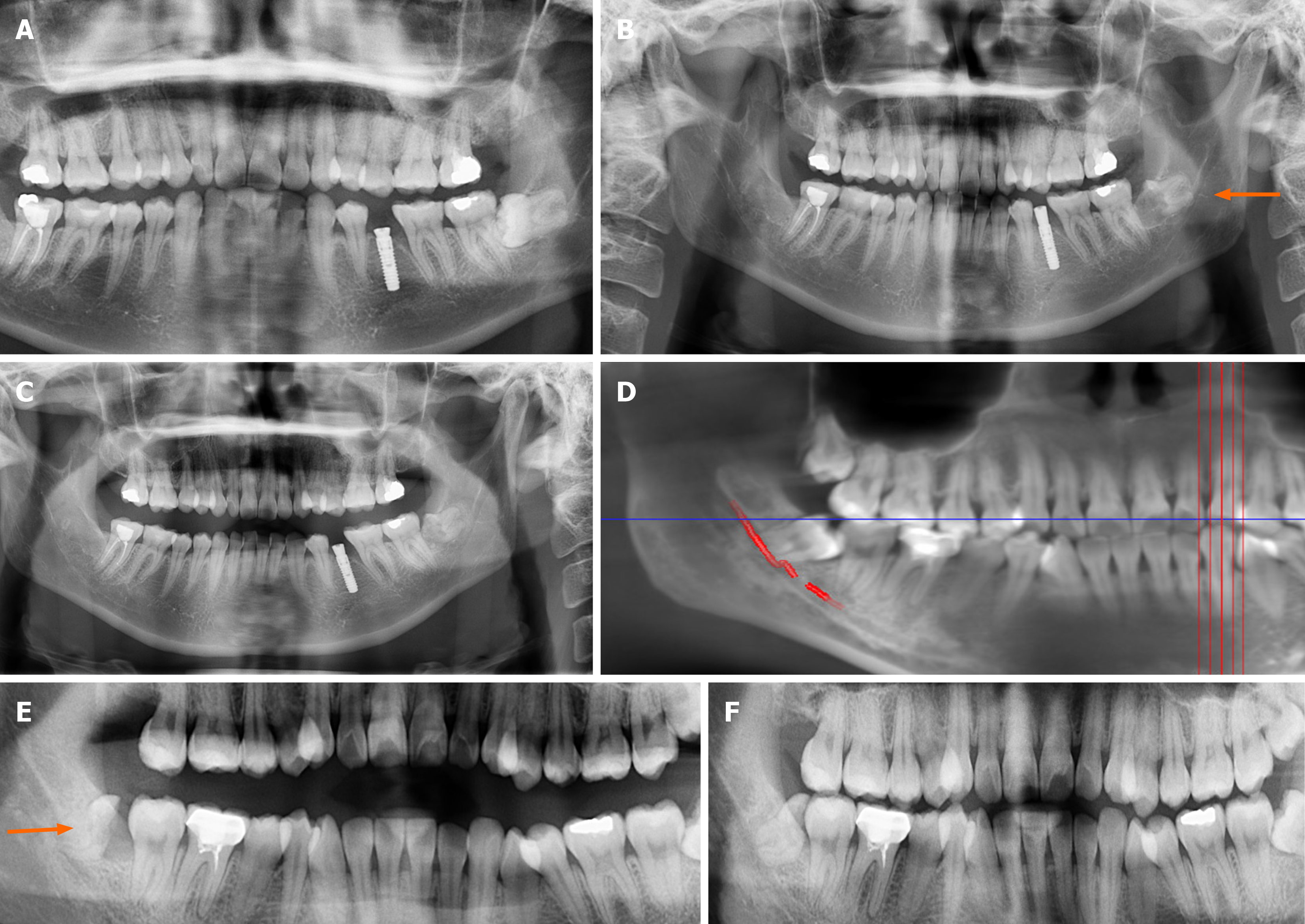
We adopted a two-stage method for IMM3 extraction. The first stage of the operation consists of grinding a major part of the IMM3 crown to obtain sufficient space for mesial movement of the IMM3. After 6-12 months, when there is distance between the root tips of the remaining part of the IMM3 and the IAN, IMM3 can be completely extracted (Figure 1).
CBCT is expensive and only used in larger dental institutions, and can only clarify the three-dimensional relationship between IMM3 and the IAN. However, if IMM3-IAN is close or IMM3 passes through IAN, routine removal of IMM3 cannot avoid postoperative complications. This approach avoids damage to the IAN. We believe that this method is worth popularizing, especially in grassroots hospitals, which may only have the capability for dental radiography and not CBCT.
Partial IMM3 grinding may be a good alternative treatment option to avoid IAN injury in high-risk cases.
The conventional method of extracting impacted mandibular third molars (IMM3) that are closely related to the inferior alveolar nerve (IAN) can easily damage the nerve.
To avoid damaging the IAN during tooth extraction.
To proposes a method for the partial grinding of an IMM3 near the IAN to prevent IAN injury during IMM3 extraction.
The first stage was to use a high-speed turbo drill to grind and cut most of the IMM3 dental crowns, leaving the roots in place. After 6-12 months, when the IMM3 root left the nerve canal, complete extraction of the IMM3 root was performed.
Although it seemed to take longer after two stages, all IMM3s were completely removed, and there were no cases of complications of damaging the IAN.
Two-stage extraction of IMM3 located closer to the IAN canal can minimize nerve damage to the greatest extent possible.
Partial IMM3 grinding may be a good alternative treatment option to avoid IAN injury in high-risk cases.
Provenance and peer review: Unsolicited article; Externally peer reviewed.
Peer-review model: Single blind
Specialty type: Medicine, research and experimental
Country/Territory of origin: China
Peer-review report’s scientific quality classification
Grade A (Excellent): 0
Grade B (Very good): 0
Grade C (Good): C
Grade D (Fair): 0
Grade E (Poor): 0
P-Reviewer: Pameijer CH, United States S-Editor: Qu XL L-Editor: Wang TQ P-Editor: Xu ZH
| 1. | Passi D, Singh G, Dutta S, Srivastava D, Chandra L, Mishra S, Srivastava A, Dubey M. Study of pattern and prevalence of mandibular impacted third molar among Delhi-National Capital Region population with newer proposed classification of mandibular impacted third molar: A retrospective study. Natl J Maxillofac Surg. 2019;10:59-67. [RCA] [PubMed] [DOI] [Full Text] [Full Text (PDF)] [Cited by in Crossref: 5] [Cited by in RCA: 29] [Article Influence: 4.8] [Reference Citation Analysis (0)] |
| 2. | Cetira Filho EL, Sales PHH, Rebelo HL, Silva PGB, Maffìa F, Vellone V, Cascone P, Leão JC, Costa FWG. Do lower third molars increase the risk of complications during mandibular sagittal split osteotomy? Systematic review and meta-analysis. Int J Oral Maxillofac Surg. 2022;51:906-921. [RCA] [PubMed] [DOI] [Full Text] [Cited by in RCA: 1] [Reference Citation Analysis (0)] |
| 3. | Sifuentes-Cervantes JS, Carrillo-Morales F, Castro-Núñez J, Cunningham LL, Van Sickels JE. Third molar surgery: Past, present, and the future. Oral Surg Oral Med Oral Pathol Oral Radiol. 2021;132:523-531. [RCA] [PubMed] [DOI] [Full Text] [Cited by in Crossref: 3] [Cited by in RCA: 41] [Article Influence: 10.3] [Reference Citation Analysis (0)] |
| 4. | Reia VCB, de Toledo Telles-Araujo G, Peralta-Mamani M, Biancardi MR, Rubira CMF, Rubira-Bullen IRF. Diagnostic accuracy of CBCT compared to panoramic radiography in predicting IAN exposure: a systematic review and meta-analysis. Clin Oral Investig. 2021;25:4721-4733. [RCA] [PubMed] [DOI] [Full Text] [Cited by in Crossref: 2] [Cited by in RCA: 15] [Article Influence: 3.8] [Reference Citation Analysis (0)] |
| 5. | Uribe S. The Routine Use of 3D Imaging May Not Reduce the Risk of Injuries to the Alveolar Inferior Nerve During Third Molar Extraction. J Evid Based Dent Pract. 2019;19:89-90. [RCA] [PubMed] [DOI] [Full Text] [Cited by in Crossref: 2] [Cited by in RCA: 1] [Article Influence: 0.2] [Reference Citation Analysis (0)] |
| 6. | Fitzpatrick E, Sharma V, Rojoa D, Raheman F, Singh H. The use of cone-beam computed tomography (CBCT) in radiocarpal fractures: a diagnostic test accuracy meta-analysis. Skeletal Radiol. 2022;51:923-934. [RCA] [PubMed] [DOI] [Full Text] [Full Text (PDF)] [Cited by in Crossref: 1] [Cited by in RCA: 10] [Article Influence: 3.3] [Reference Citation Analysis (0)] |
| 7. | Klatt JC, Sorowka T, Kluwe L, Smeets R, Gosau M, Hanken H. Does a preoperative cone beam CT reduce complication rates in the surgical removal of complex lower third molars? A retrospective study including 486 cases. Head Face Med. 2021;17:33. [RCA] [PubMed] [DOI] [Full Text] [Full Text (PDF)] [Cited by in Crossref: 3] [Cited by in RCA: 2] [Article Influence: 0.5] [Reference Citation Analysis (0)] |
| 8. | Hasani M, Razavi N, Haghnegahdar A, Zarifi M. Evaluating the presence of IAN injury in patients with juxta-apical radiolucency after third molar surgery: a retrospective cohort study. BMC Oral Health. 2021;21:428. [RCA] [PubMed] [DOI] [Full Text] [Full Text (PDF)] [Reference Citation Analysis (0)] |
| 9. | Bhat P, Cariappa KM. Inferior alveolar nerve deficits and recovery following surgical removal of impacted mandibular third molars. J Maxillofac Oral Surg. 2012;11:304-308. [RCA] [PubMed] [DOI] [Full Text] [Cited by in Crossref: 14] [Cited by in RCA: 18] [Article Influence: 1.4] [Reference Citation Analysis (0)] |
| 10. | Kang F, Sah MK, Fei G. Determining the risk relationship associated with inferior alveolar nerve injury following removal of mandibular third molar teeth: A systematic review. J Stomatol Oral Maxillofac Surg. 2020;121:63-69. [RCA] [PubMed] [DOI] [Full Text] [Cited by in Crossref: 16] [Cited by in RCA: 17] [Article Influence: 3.4] [Reference Citation Analysis (0)] |
| 11. | Daware SN, Balakrishna R, Deogade SC, Ingole YS, Patil SM, Naitam DM. Assessment of postoperative discomfort and nerve injuries after surgical removal of mandibular third molar: A prospective study. J Family Med Prim Care. 2021;10:1712-1717. [RCA] [PubMed] [DOI] [Full Text] [Full Text (PDF)] [Cited by in Crossref: 2] [Cited by in RCA: 9] [Article Influence: 2.3] [Reference Citation Analysis (0)] |
| 12. | Bozkurt P, Görürgöz C. Detecting direct inferior alveolar nerve - Third molar contact and canal decorticalization by cone-beam computed tomography to predict postoperative sensory impairment. J Stomatol Oral Maxillofac Surg. 2020;121:259-263. [RCA] [PubMed] [DOI] [Full Text] [Cited by in Crossref: 3] [Cited by in RCA: 2] [Article Influence: 0.3] [Reference Citation Analysis (0)] |
| 13. | Hoshi R, Tetsumura A, Yamaguchi S. Preoperative imaging findings as predictors of postoperative inferior alveolar nerve injury following mandibular cyst surgery. J Oral Sci. 2018;60:618-625. [RCA] [PubMed] [DOI] [Full Text] [Cited by in Crossref: 1] [Cited by in RCA: 1] [Article Influence: 0.2] [Reference Citation Analysis (0)] |
| 14. | Feher B, Spandl LF, Lettner S, Ulm C, Gruber R, Kuchler U. Prediction of post-traumatic neuropathy following impacted mandibular third molar removal. J Dent. 2021;115:103838. [RCA] [PubMed] [DOI] [Full Text] [Reference Citation Analysis (0)] |
| 15. | Ali AS, Benton JA, Yates JM. Risk of inferior alveolar nerve injury with coronectomy vs surgical extraction of mandibular third molars-A comparison of two techniques and review of the literature. J Oral Rehabil. 2018;45:250-257. [RCA] [PubMed] [DOI] [Full Text] [Cited by in Crossref: 18] [Cited by in RCA: 33] [Article Influence: 4.1] [Reference Citation Analysis (0)] |
| 16. | Pitros P, O'Connor N, Tryfonos A, Lopes V. A systematic review of the complications of high-risk third molar removal and coronectomy: development of a decision tree model and preliminary health economic analysis to assist in treatment planning. Br J Oral Maxillofac Surg. 2020;58:e16-e24. [RCA] [PubMed] [DOI] [Full Text] [Cited by in Crossref: 8] [Cited by in RCA: 21] [Article Influence: 4.2] [Reference Citation Analysis (0)] |
| 17. | Kuntz NM, Schulze R. Three-Dimensional Classification of Lower Third Molars and Their Relationship to the Mandibular Canal. J Oral Maxillofac Surg. 2021;79:1611-1620. [RCA] [PubMed] [DOI] [Full Text] [Cited by in Crossref: 3] [Cited by in RCA: 3] [Article Influence: 0.8] [Reference Citation Analysis (0)] |
| 18. | Lampert RC, Nesbitt TR, Chuang SK, Ziccardi VB. Management of endodontic injuries to the inferior alveolar nerve. Quintessence Int. 2016;47:581-587. [RCA] [PubMed] [DOI] [Full Text] [Cited by in RCA: 1] [Reference Citation Analysis (0)] |
| 19. | Caughey JA, Do Q, Shen D, Ohyama H, He P, Tubbs RS, Iwanaga J. Comprehensive review of the incisive branch of the inferior alveolar nerve. Anat Cell Biol. 2021;54:409-416. [RCA] [PubMed] [DOI] [Full Text] [Full Text (PDF)] [Cited by in Crossref: 1] [Cited by in RCA: 6] [Article Influence: 1.5] [Reference Citation Analysis (0)] |
| 20. | Abu-Mostafa N, AlRejaie LM, Almutairi FA, Alajaji RA, Alkodair MM, Alzahem NA. Evaluation of the Outcomes of Coronectomy Procedure versus Surgical Extraction of Lower Third Molars Which Have a High Risk for Inferior Alveolar Nerve Injury: A Systematic Review. Int J Dent. 2021;2021:9161606. [RCA] [PubMed] [DOI] [Full Text] [Full Text (PDF)] [Cited by in Crossref: 1] [Cited by in RCA: 8] [Article Influence: 2.0] [Reference Citation Analysis (0)] |


