Copyright
©The Author(s) 2021.
World J Clin Cases. Mar 16, 2021; 9(8): 1885-1892
Published online Mar 16, 2021. doi: 10.12998/wjcc.v9.i8.1885
Published online Mar 16, 2021. doi: 10.12998/wjcc.v9.i8.1885
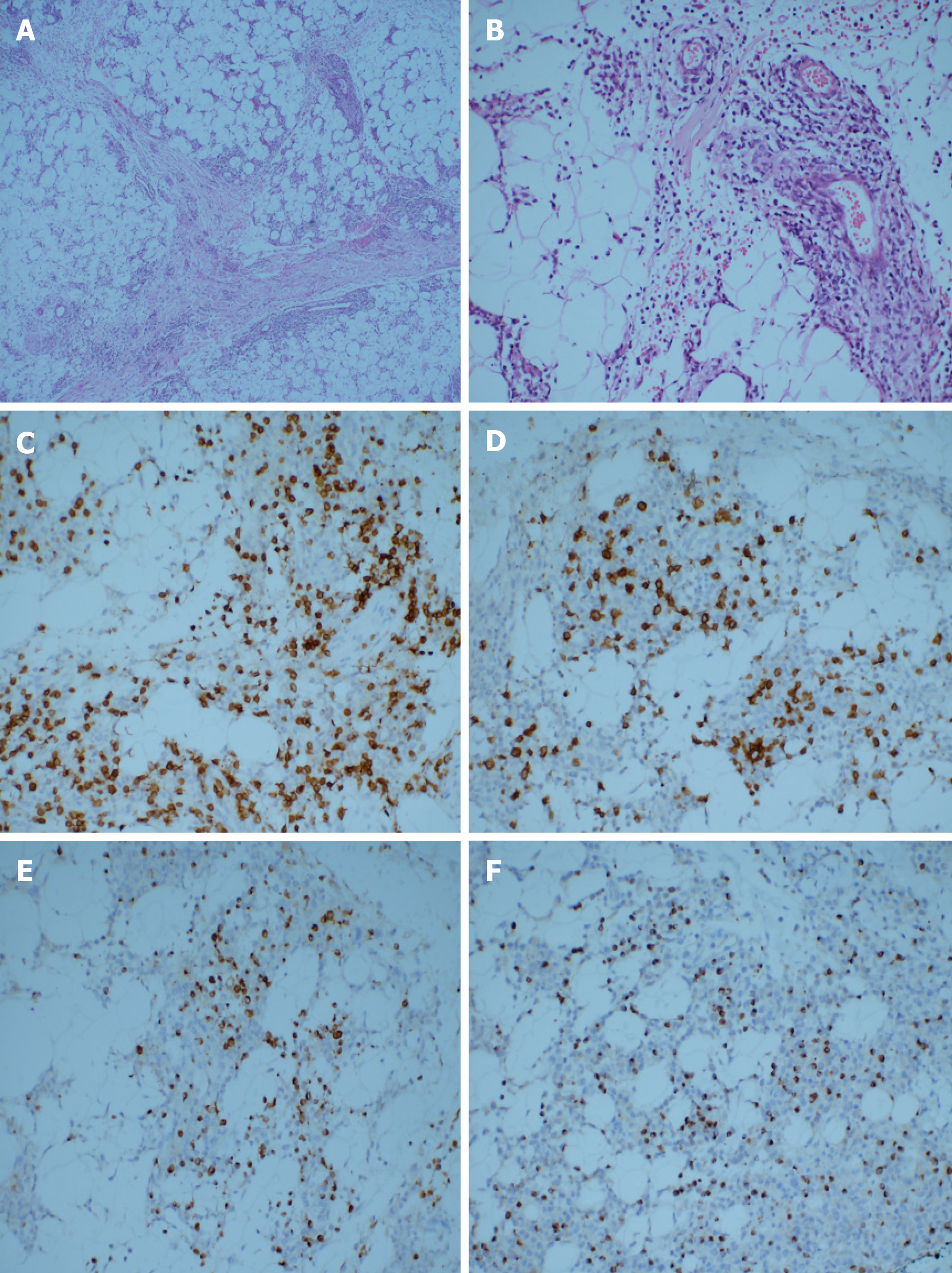
Figure 1 Pathological examination.
A and B: Hematoxylin-eosin staining. A large number of lymphocytes infiltrated the fibrous connective tissue and adipose tissue. Some cells were characterized by abundant cytoplasm and hyperchromatic nuclei with obvious atypia. The infiltrating cells were distributed in a typical flower-ring-like pattern around the fat cells (original magnification A: 40 ×; B: 200 ×); C-F: Immunohistological staining showing tumor positivity for CD3, CD8, Gram-B, and T-cell restricted intracellular antigen-1 (original magnification 200 ×); C: CD3; D: CD8; E: Gram-B; F: T-cell restricted intracellular antigen-1.
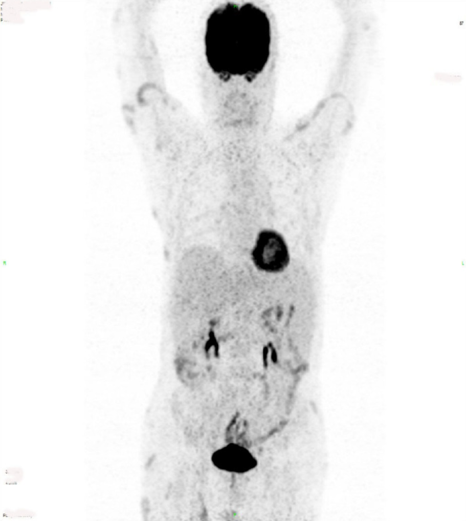
Figure 2 Multiple nodular and patchy radiation uptake increased shadows were found in the bilateral upper arms, left chest wall, bilateral abdominal wall, bilateral buttocks, and bilateral subcutaneous thighs.
The maximum standard uptake value was 5.1. No obvious abnormality was found in the distribution of skeletal radioactivity.
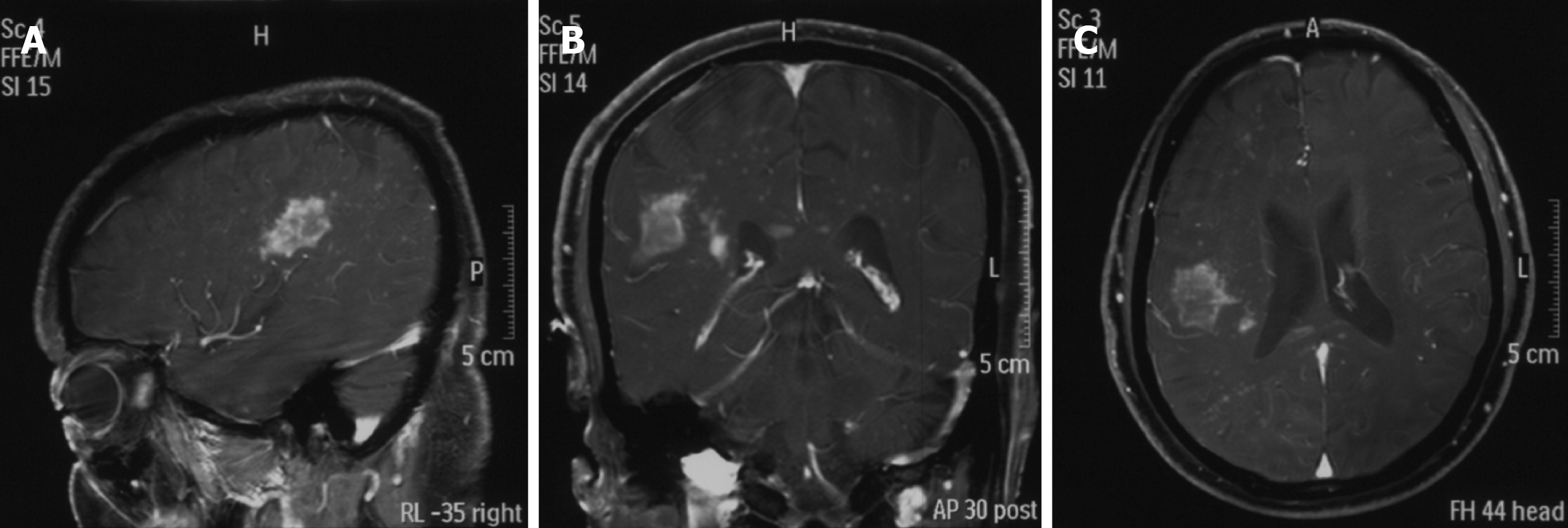
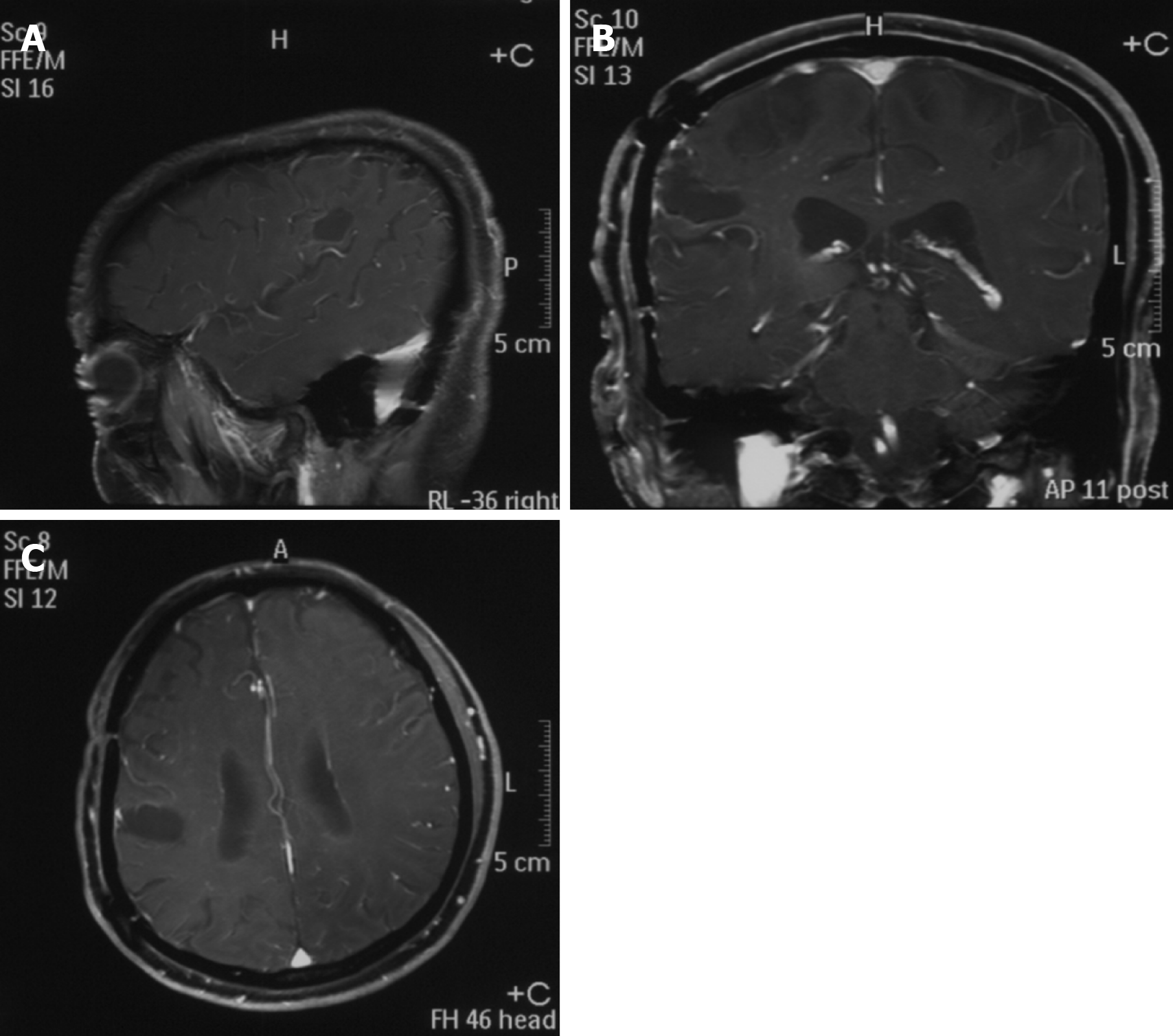
Figure 3 Preoperative magnetic resonance imaging revealed a remarkably enhanced mass.
A: Sagittal enhanced imaging lesion in the region of the right temporal lobe; B: Coronal enhanced imaging lesion in the region of the right temporal lobe; C: Axial enhanced imaging lesion in the region of the right temporal lobe.
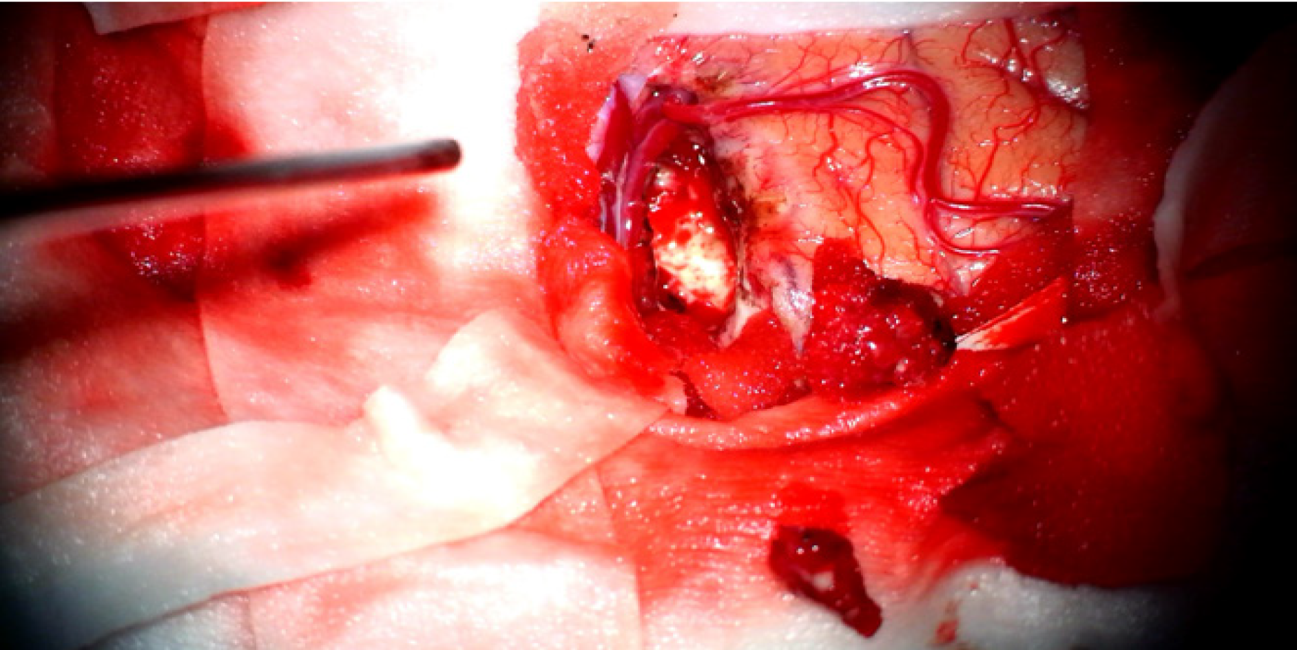
Figure 4 The lesion was gray-white in color, and soft tissue with rich blood supply blurred the boundary with the surrounding brain tissue.
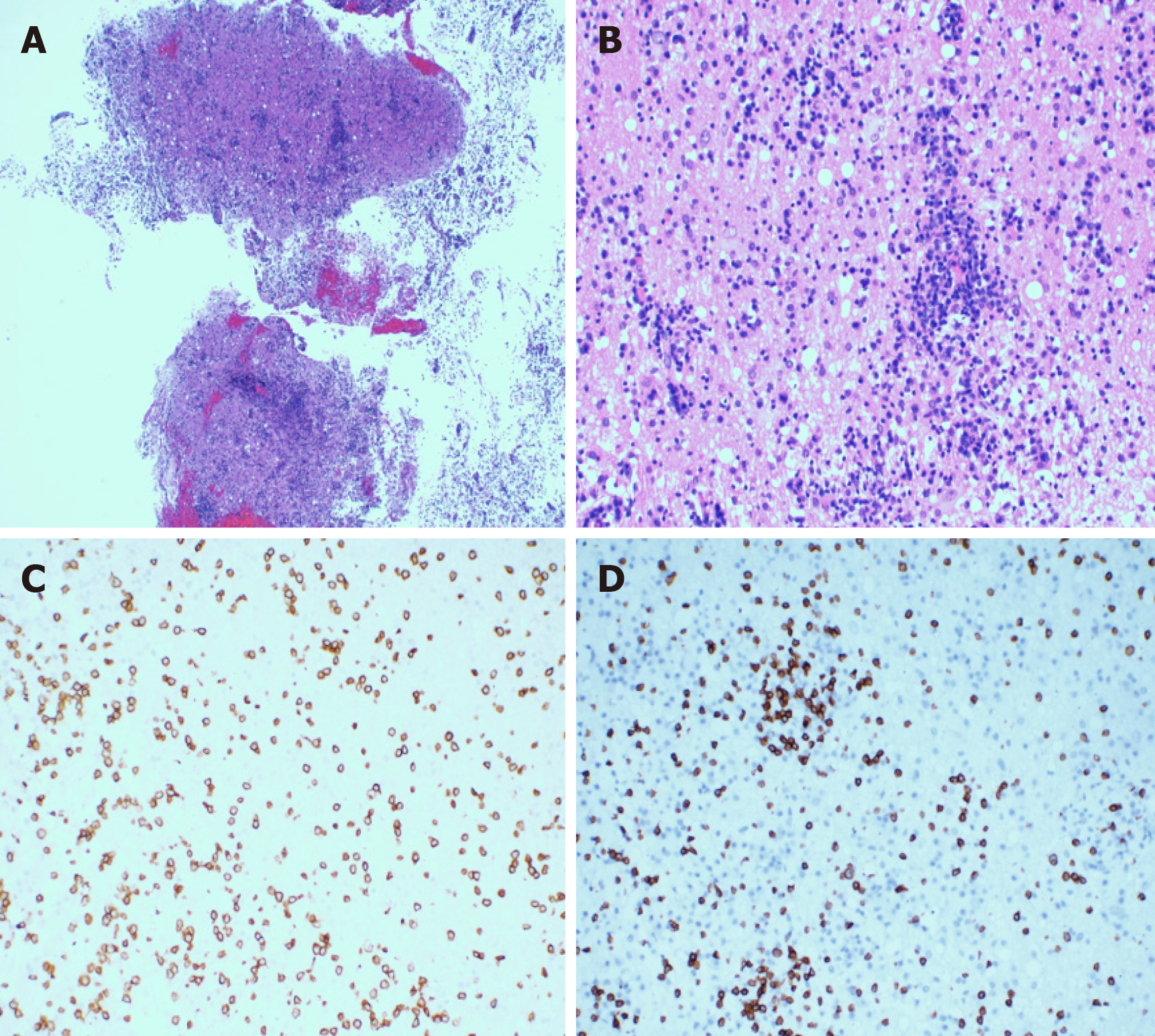
Figure 5 The hematoxylin-eosin staining of the resected specimens revealed that the plasmocytes and lymphocytes had proliferated.
A and B: The lymphocytes infiltrated the blood vessels and showed a vascular-centered growth pattern (A: 40 ×; B: 200 ×); C and D: Immunohistological staining showing the intracranial tumor positivity for CD3 and CD8 (original magnification 200 ×); C: CD3; D: CD8.
Figure 6 Nine months after the craniocerebral surgery, brain magnetic resonance imaging showed no sign of recurrence of the tumor.
A: Sagittal enhanced imaging; B: Coronal enhanced imaging; C: Axial enhanced imaging.
- Citation: Sun J, Ma XS, Qu LM, Song XS. Subcutaneous panniculitis-like T-cell lymphoma invading central nervous system in long-term clinical remission with lenalidomide: A case report. World J Clin Cases 2021; 9(8): 1885-1892
- URL: https://www.wjgnet.com/2307-8960/full/v9/i8/1885.htm
- DOI: https://dx.doi.org/10.12998/wjcc.v9.i8.1885


