Copyright
©The Author(s) 2021.
World J Clin Cases. Mar 16, 2021; 9(8): 1885-1892
Published online Mar 16, 2021. doi: 10.12998/wjcc.v9.i8.1885
Published online Mar 16, 2021. doi: 10.12998/wjcc.v9.i8.1885
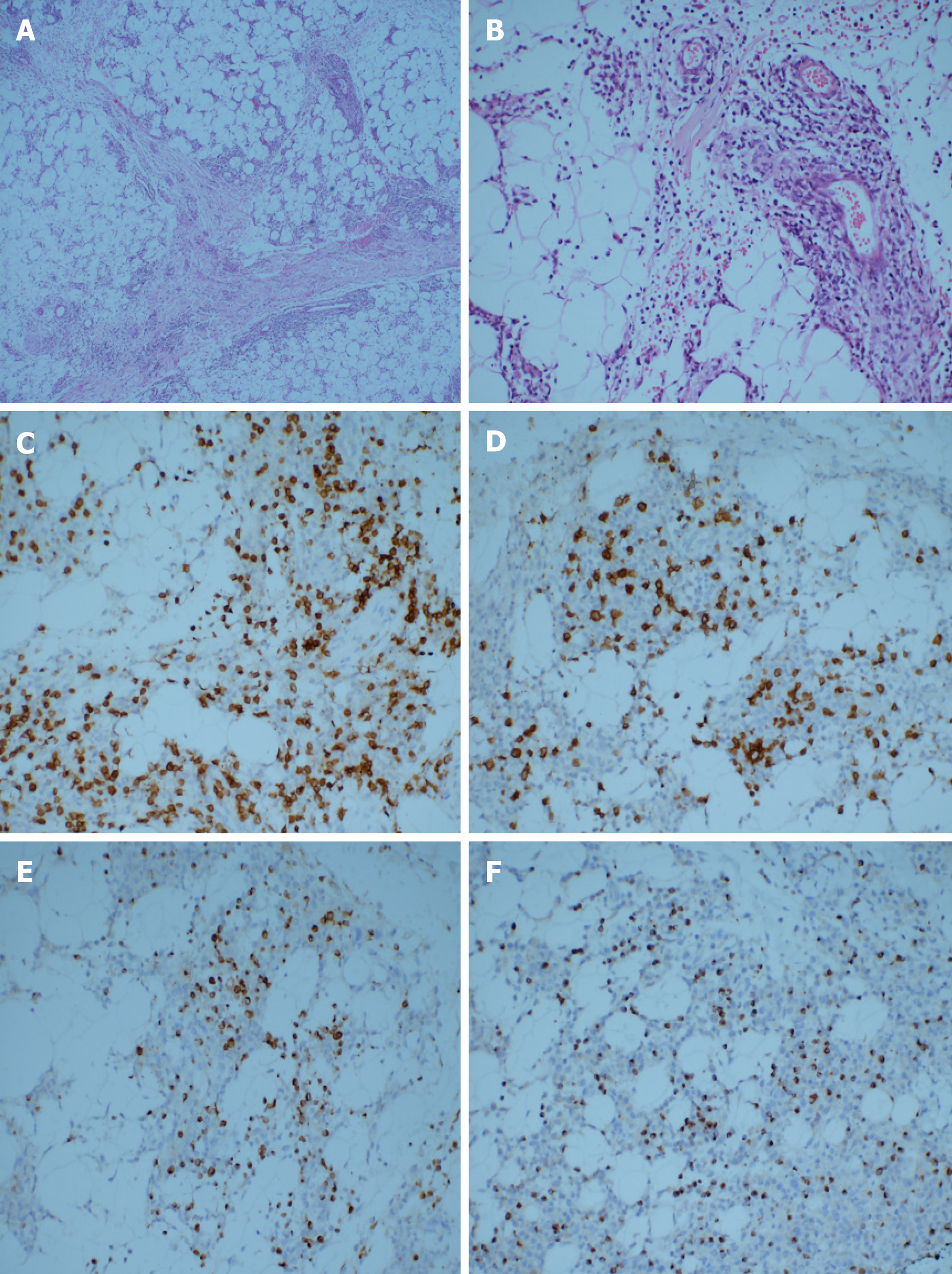
Figure 1 Pathological examination.
A and B: Hematoxylin-eosin staining. A large number of lymphocytes infiltrated the fibrous connective tissue and adipose tissue. Some cells were characterized by abundant cytoplasm and hyperchromatic nuclei with obvious atypia. The infiltrating cells were distributed in a typical flower-ring-like pattern around the fat cells (original magnification A: 40 ×; B: 200 ×); C-F: Immunohistological staining showing tumor positivity for CD3, CD8, Gram-B, and T-cell restricted intracellular antigen-1 (original magnification 200 ×); C: CD3; D: CD8; E: Gram-B; F: T-cell restricted intracellular antigen-1.
- Citation: Sun J, Ma XS, Qu LM, Song XS. Subcutaneous panniculitis-like T-cell lymphoma invading central nervous system in long-term clinical remission with lenalidomide: A case report. World J Clin Cases 2021; 9(8): 1885-1892
- URL: https://www.wjgnet.com/2307-8960/full/v9/i8/1885.htm
- DOI: https://dx.doi.org/10.12998/wjcc.v9.i8.1885


