Copyright
©The Author(s) 2020.
World J Clin Cases. Oct 6, 2020; 8(19): 4644-4651
Published online Oct 6, 2020. doi: 10.12998/wjcc.v8.i19.4644
Published online Oct 6, 2020. doi: 10.12998/wjcc.v8.i19.4644
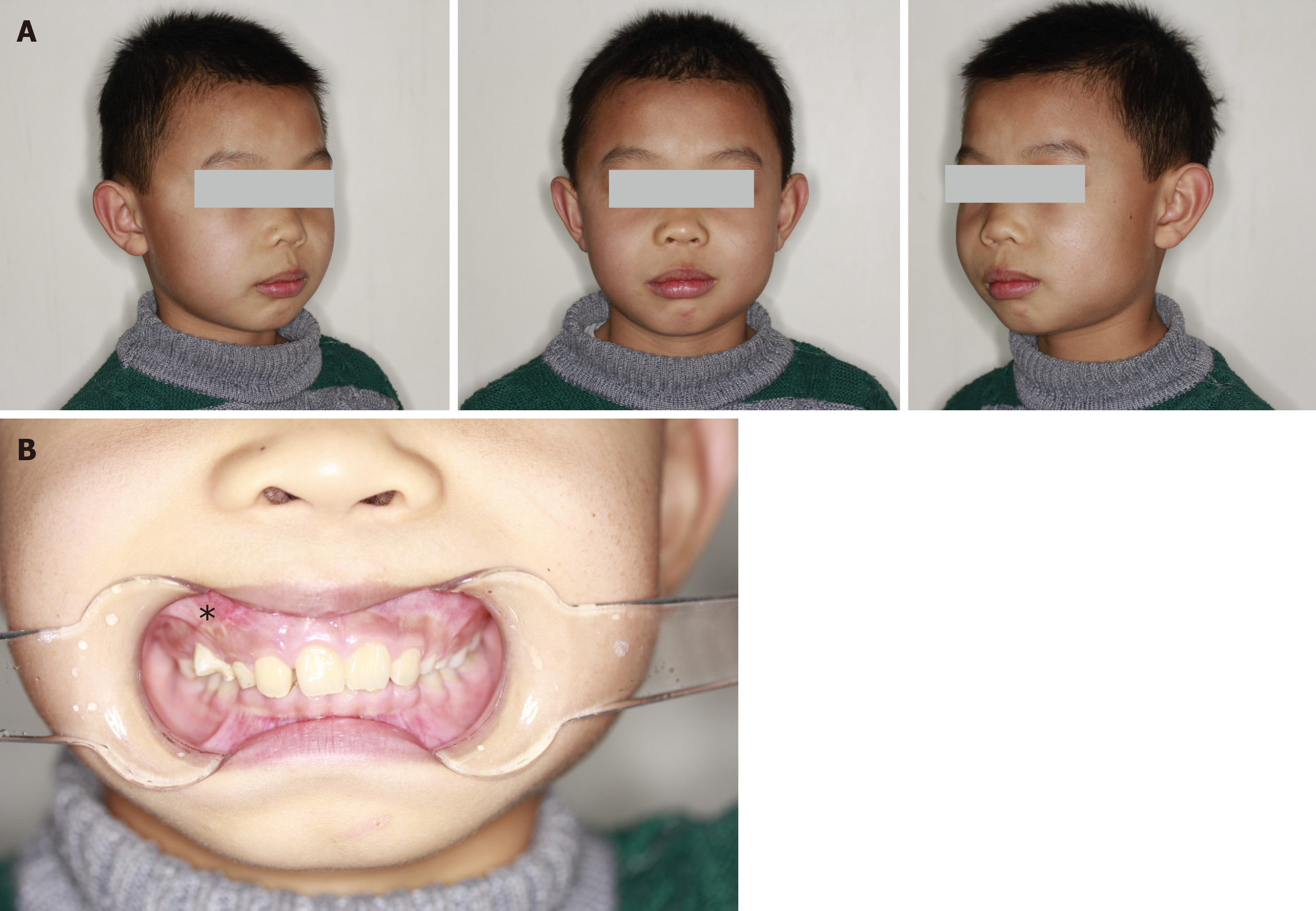
Figure 1 Physical examination of the maxillofacial region.
A: The face was symmetrical; B: The upper right side of the vestibular groove was shoaled.
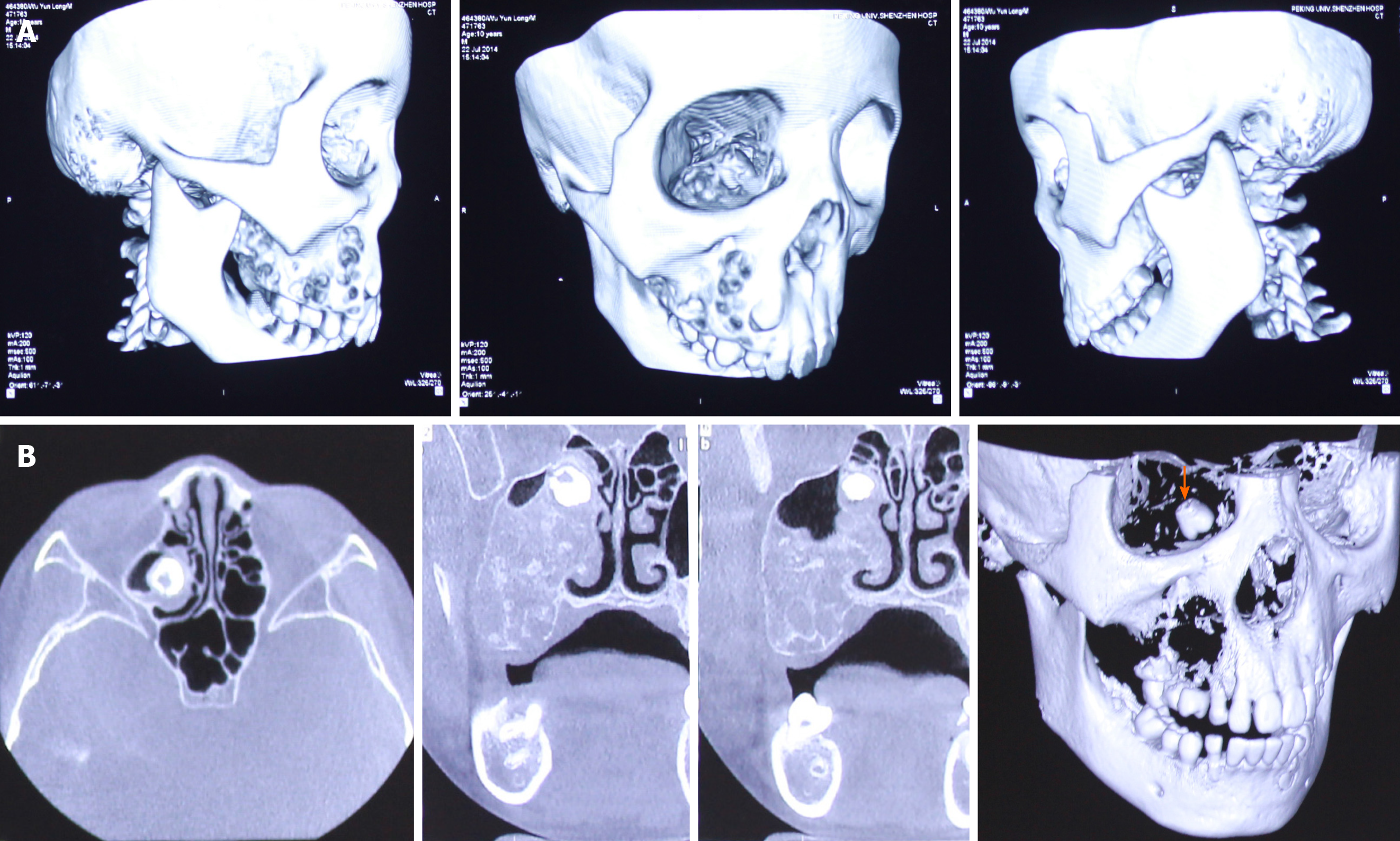
Figure 2 Computed tomography scan and 3D reconstruction displayed a multicystic low-density shadow in the right maxilla.
A: The body of the right maxilla manifested irregular swelling; B: The orange arrow shows a molar that was squeezed into the right maxillary sinus and embedded in the superior part of the lesion close to the canalis opticus.
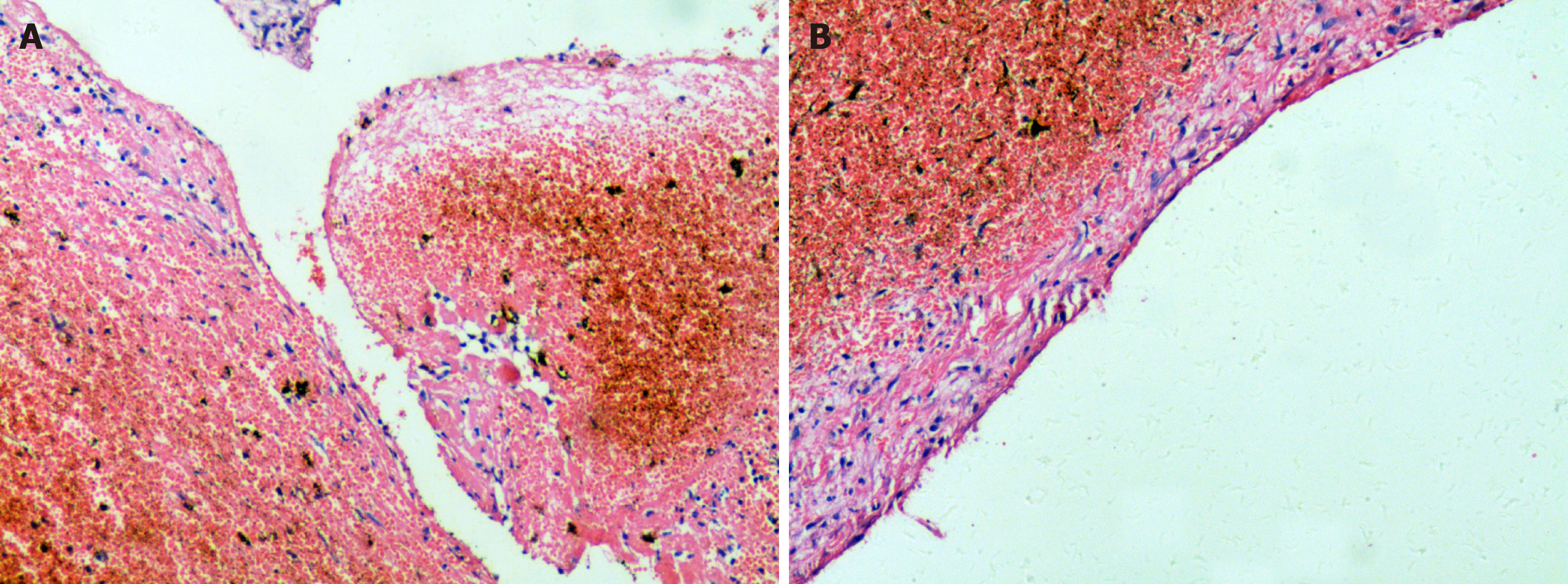
Figure 3 Microscopic examination with haematoxylin and eosin staining was performed.
A: No signs of epithelium were found, although a fibrous tissue lining was observed; B: Remote hemorrhage and clot organization supported the diagnosis of hemophilic pseudotumor (magnification, × 100).
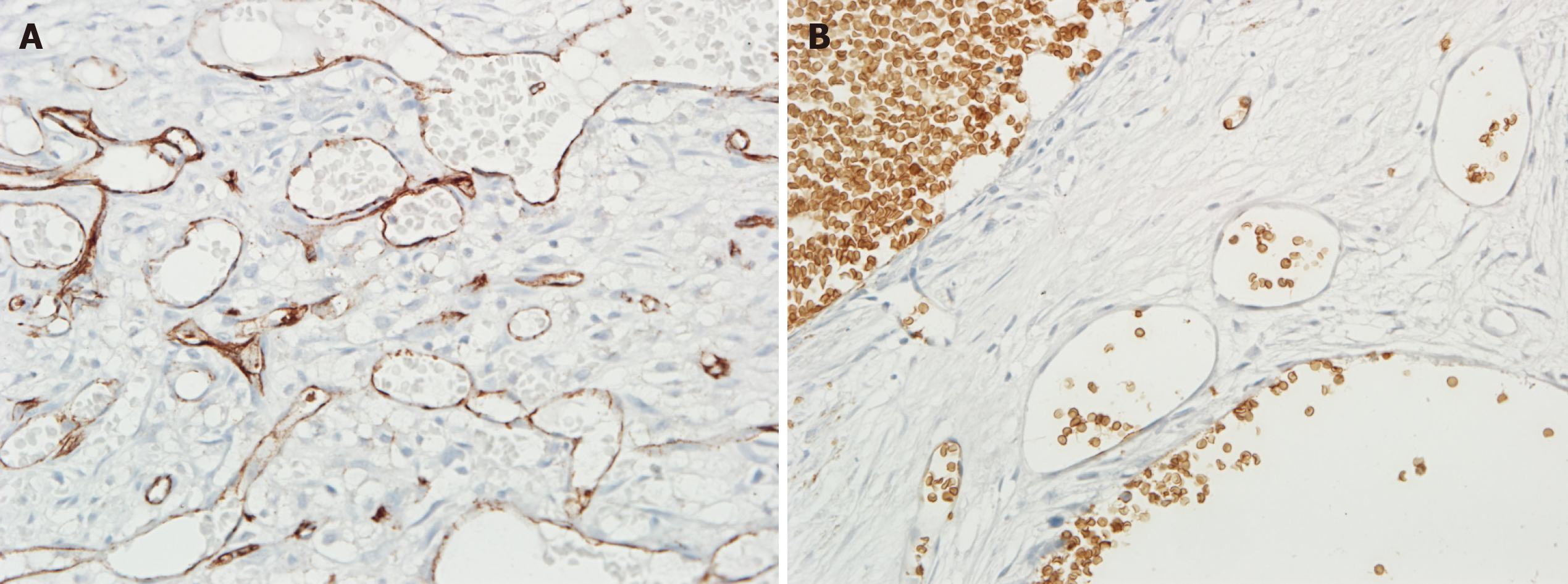
Figure 4 Immunohistochemical findings of the lesion of the second surgery.
A: Immunohistochemistry results revealed that the lesions of the second surgery were abundant in the vessels positive for CD31, a marker of endothelial components; B: The vessel endothelium of the lesions was negative for GLUT-1 (magnification, × 200).
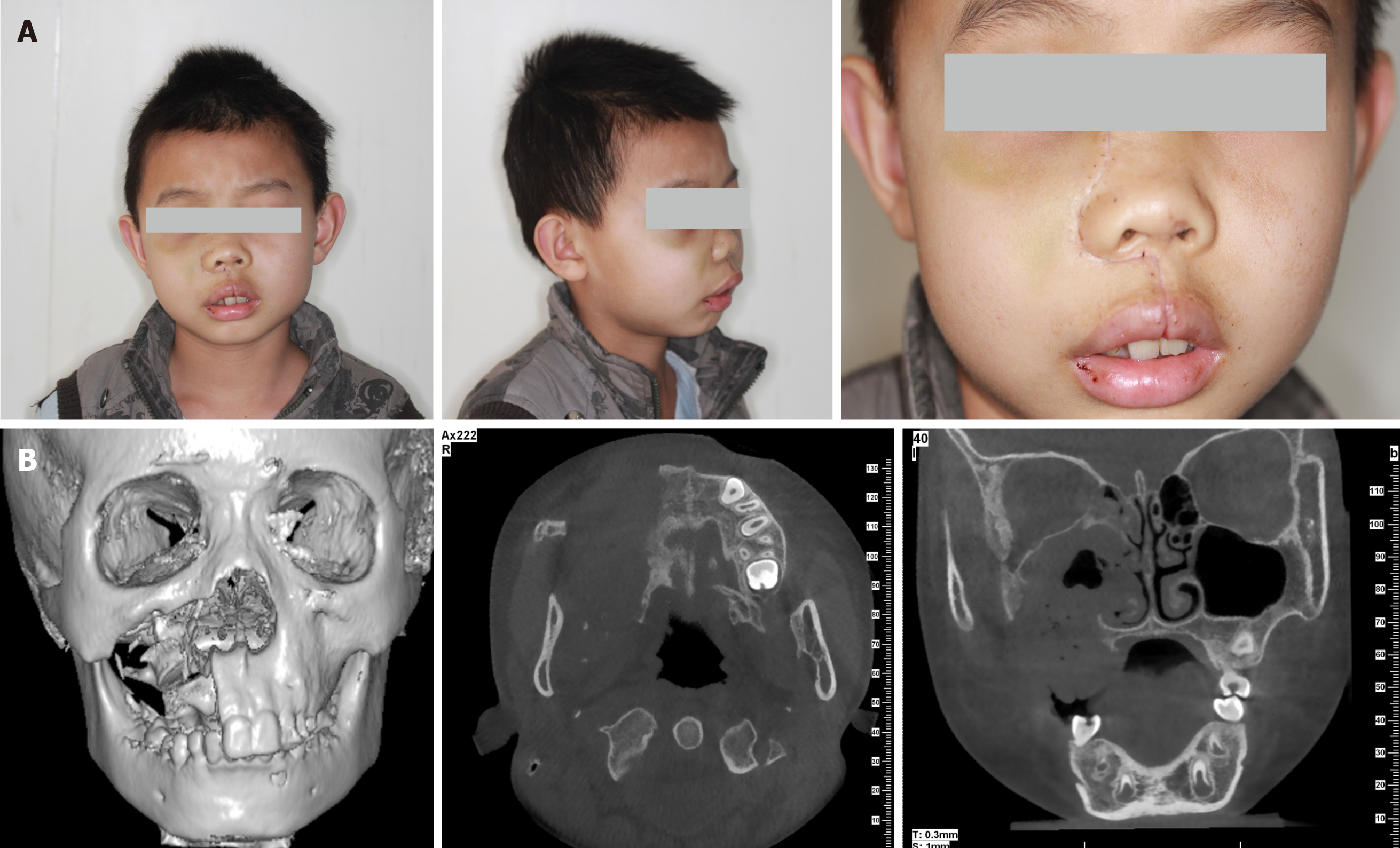
Figure 5 Physical examination of the maxillofacial region half a month after surgery.
A: The faciomaxillary region was supported by the residual bone and the face was almost symmetrical; B: Computed tomography scan and 3D reconstruction displayed complete resection of the lesion of the maxilla and removal of the contents of the maxillary sinus and ethmoidal cellules. The pavimentum orbitae and zygomatic process of the maxilla were retained.
- Citation: Cai X, Yu JJ, Tian H, Shan ZF, Liu XY, Jia J. Intraosseous venous malformation of the maxilla after enucleation of a hemophilic pseudotumor: A case report. World J Clin Cases 2020; 8(19): 4644-4651
- URL: https://www.wjgnet.com/2307-8960/full/v8/i19/4644.htm
- DOI: https://dx.doi.org/10.12998/wjcc.v8.i19.4644


