Copyright
©The Author(s) 2020.
World J Clin Cases. Oct 6, 2020; 8(19): 4624-4632
Published online Oct 6, 2020. doi: 10.12998/wjcc.v8.i19.4624
Published online Oct 6, 2020. doi: 10.12998/wjcc.v8.i19.4624
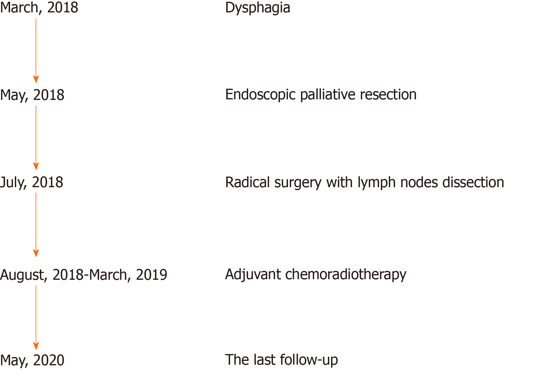
Figure 1 Timeline of the patient with esophageal carcinosarcoma.
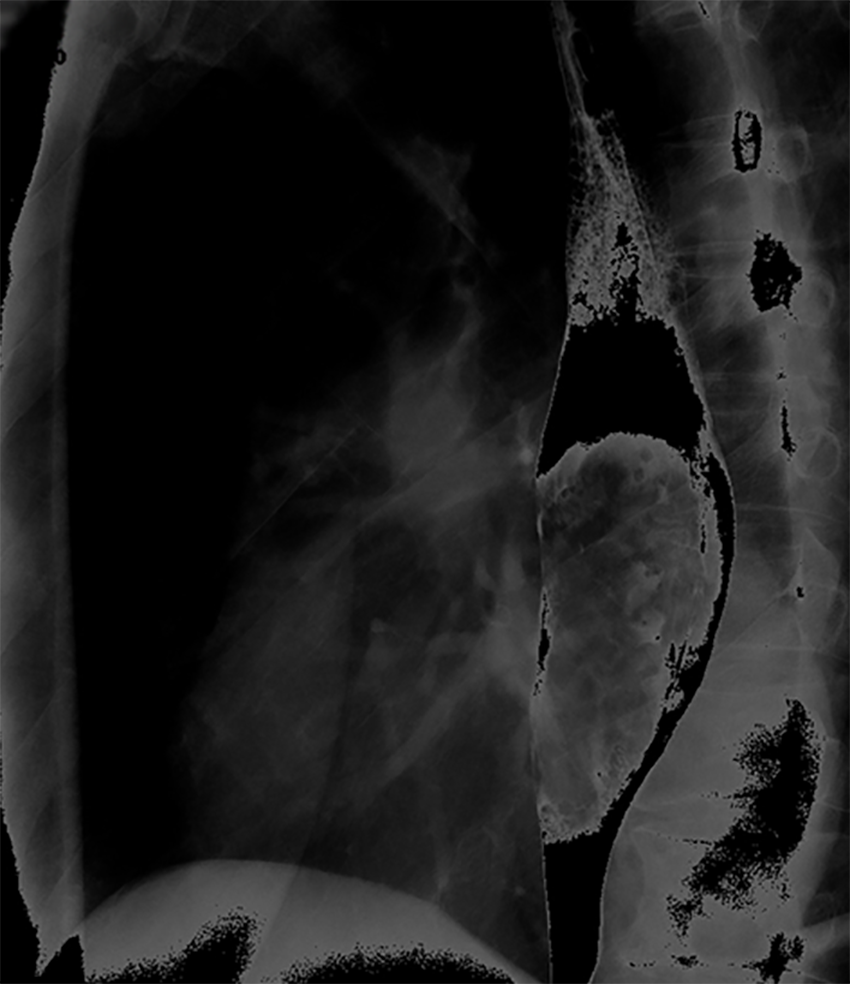
Figure 2 X-ray barium meal examination showing a huge intraluminal stalk-like mass along the middle and lower esophagus.
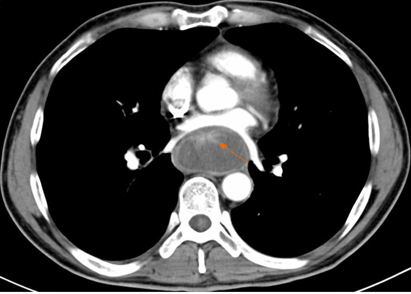
Figure 3 Computed tomography showing a prominently enhanced anterior area of the giant mass beside the esophageal wall (orange arrow), and the maximum sectional area was about 5.
6 cm × 3.5 cm.
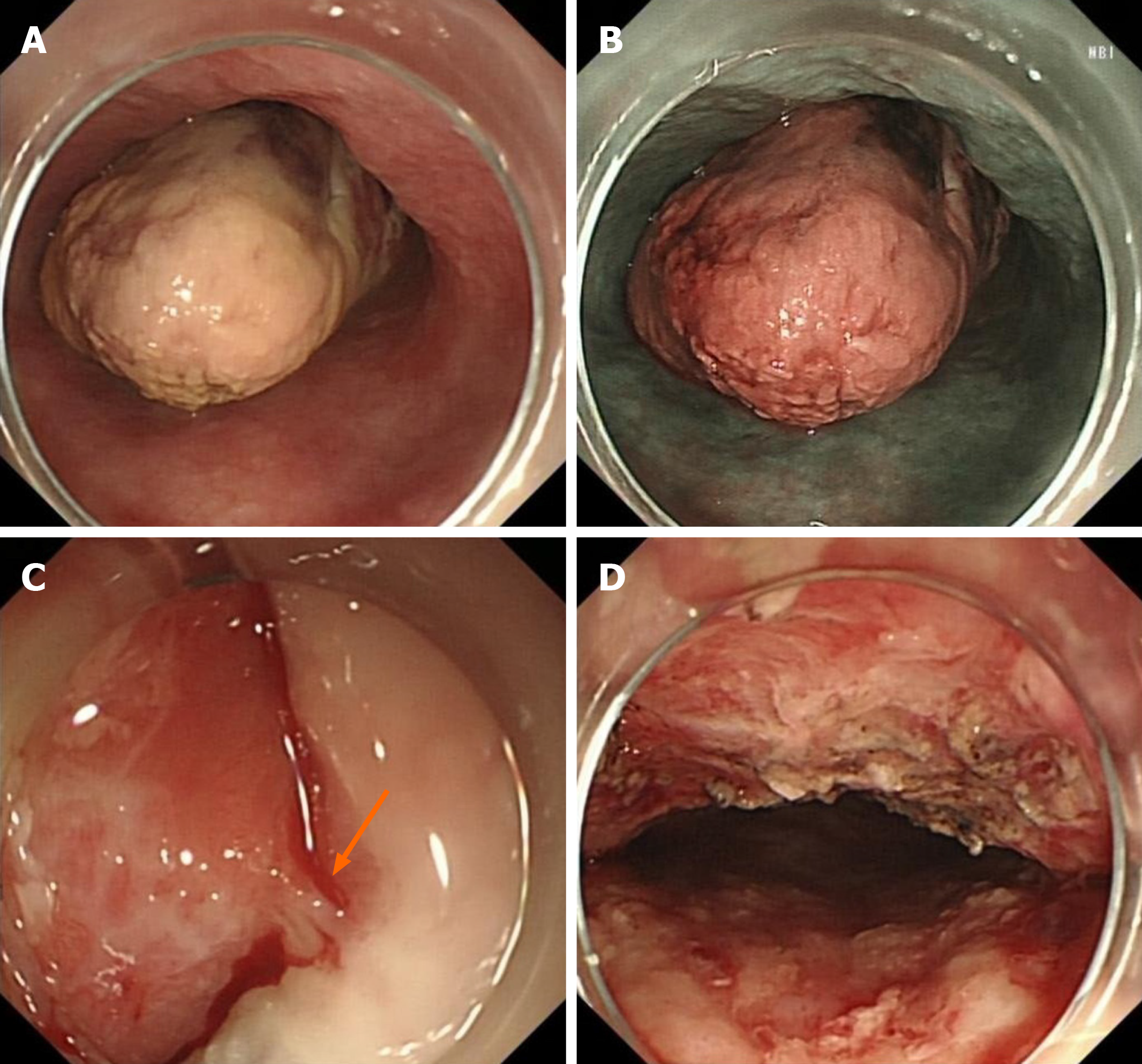
Figure 4 Endoscopy.
A: The giant mass was polypoid, gray-white, almost filling the whole esophageal lumen (white light), but the endoscope could still pass through; B: The giant polypoid mass was brown, as revealed by narrow-band imaging; C: The bulky pedicle of the mass (orange arrow); D: The wound was intact after cutting off the giant mass.
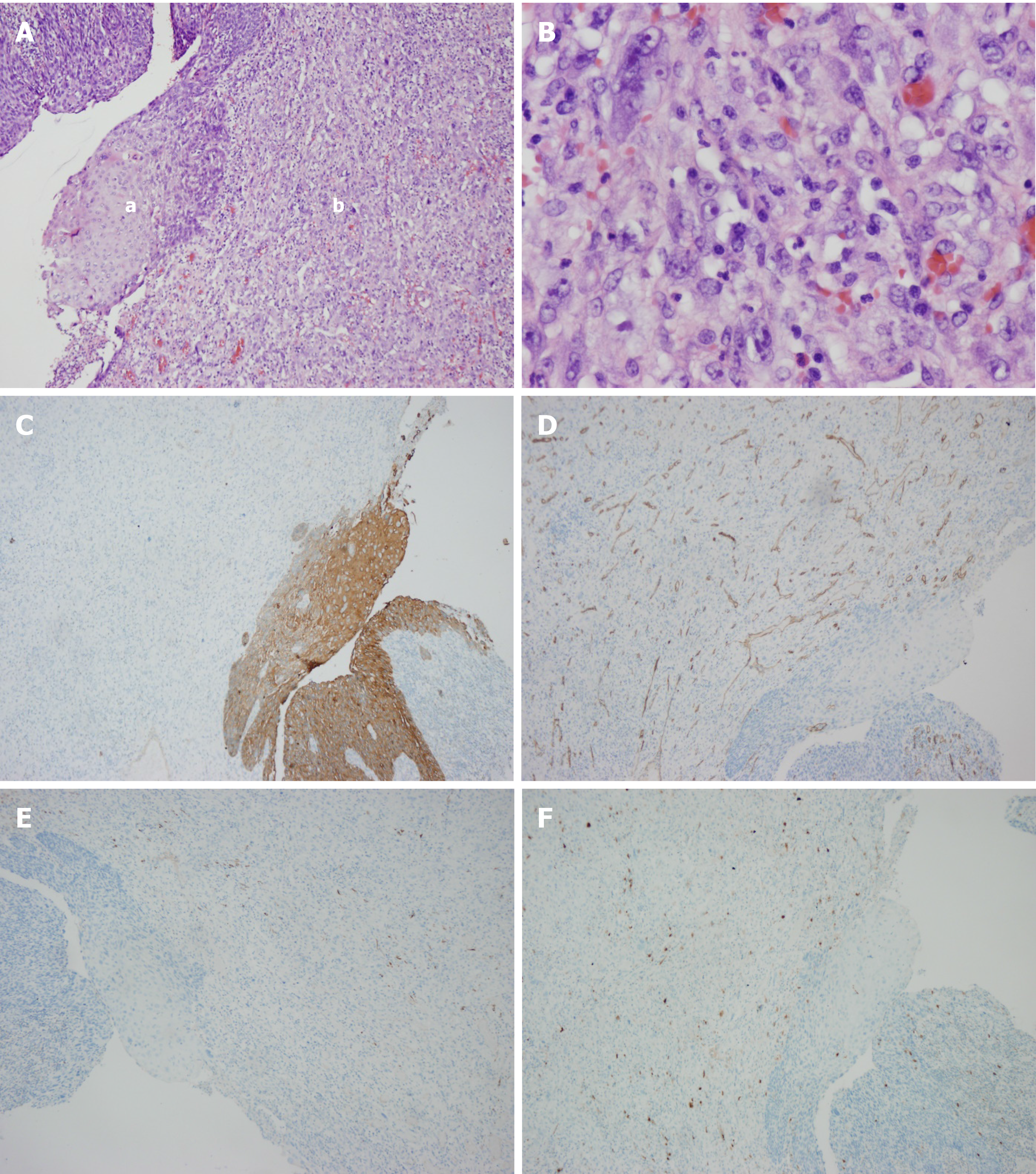
Figure 5 Postoperative pathology.
A: Hematoxylin–eosin staining showing esophageal squamous cell carcinoma (a) with sarcomatoid component (b); B: Magnification of the sarcomatoid component; C: PCK staining; D: CD34 staining; E: Desmin staining; F: S-100 staining.
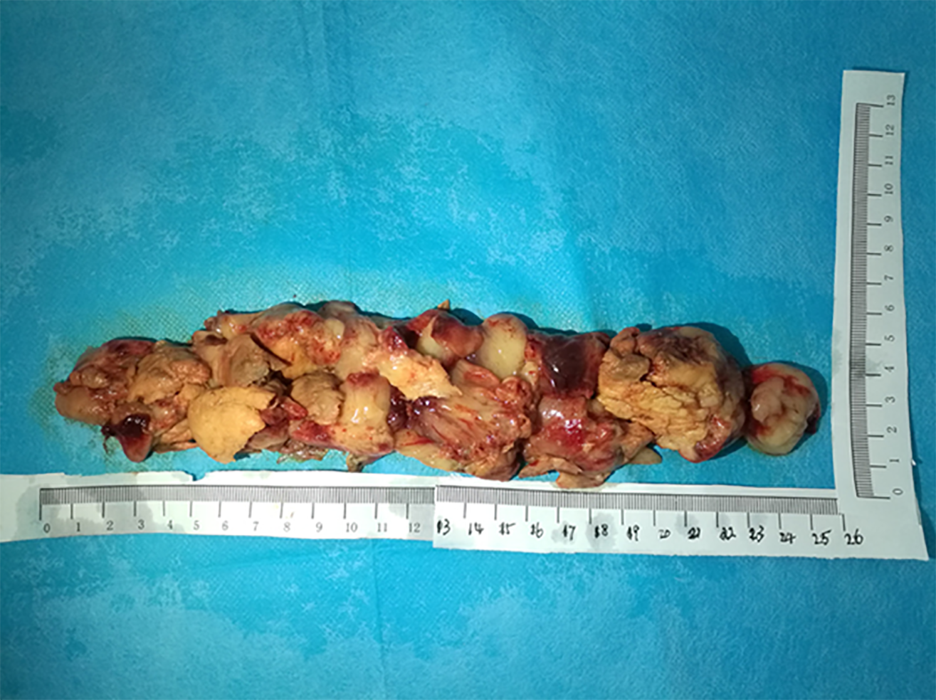
Figure 6 The giant mass was assembled after pulling out and was about 26 cm × 5 cm × 4 cm in size.
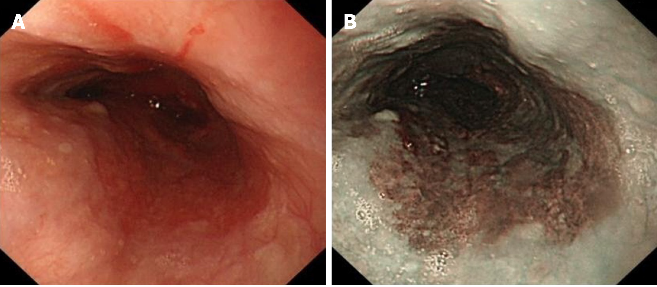
Figure 7 Endoscopic reexamination after 1 mo revealed no esophageal obstruction.
A: White light; B: Narrow-band imaging.
- Citation: Li Y, Guo LJ, Ma YC, Ye LS, Hu B. Endoscopic palliative resection of a giant 26-cm esophageal tumor: A case report. World J Clin Cases 2020; 8(19): 4624-4632
- URL: https://www.wjgnet.com/2307-8960/full/v8/i19/4624.htm
- DOI: https://dx.doi.org/10.12998/wjcc.v8.i19.4624


