Copyright
©The Author(s) 2020.
World J Clin Cases. Oct 6, 2020; 8(19): 4609-4614
Published online Oct 6, 2020. doi: 10.12998/wjcc.v8.i19.4609
Published online Oct 6, 2020. doi: 10.12998/wjcc.v8.i19.4609
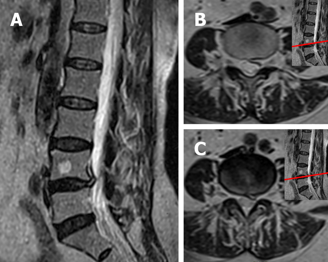
Figure 1 Magnetic resonance imaging obtained at the patient’s initial visit.
A: Sagittal T2-weighted magnetic resonance imaging revealed an L4/5 lumbar disk herniation moved upward to above the L4/5 disc level, lying behind the L4 vertebral body; B and C: Axial T2-weighted magnetic resonance imaging revealed that the lumbar disk herniation compressed the L4 nerve root at the left posterolateral of the L4 vertebral body.
Figure 2 Magnetic resonance imaging obtained 10 wk after the patient’s initial visit.
A: Sagittal T2-weighted magnetic resonance imaging revealed that most of the upwardly displaced disc material had disappeared; B and C: Axial T2-weighted magnetic resonance imaging revealed that a small amount of herniated disc material was visible at the left posterolateral of the L4 vertebral body and the L4/5 disc.
Figure 3 Magnetic resonance imaging obtained 2 years after the patient’s initial visit.
A: Sagittal T2-weighted magnetic resonance imaging (MRI) revealed the upwardly displaced disc material had almost completely disappeared; B: Axial T2-weighted MRI revealed that a small amount of herniated disc material was visible at the left posterolateral of the L4 vertebral body; and C: Axial T2-weighted MRI revealed that the herniated disc material at the left posterolateral of the L4/5 disc had disappeared.
- Citation: Wang Y, Liao SC, Dai GG, Jiang L. Resorption of upwardly displaced lumbar disk herniation after nonsurgical treatment: A case report. World J Clin Cases 2020; 8(19): 4609-4614
- URL: https://www.wjgnet.com/2307-8960/full/v8/i19/4609.htm
- DOI: https://dx.doi.org/10.12998/wjcc.v8.i19.4609


