Copyright
©The Author(s) 2020.
World J Clin Cases. Oct 6, 2020; 8(19): 4572-4578
Published online Oct 6, 2020. doi: 10.12998/wjcc.v8.i19.4572
Published online Oct 6, 2020. doi: 10.12998/wjcc.v8.i19.4572
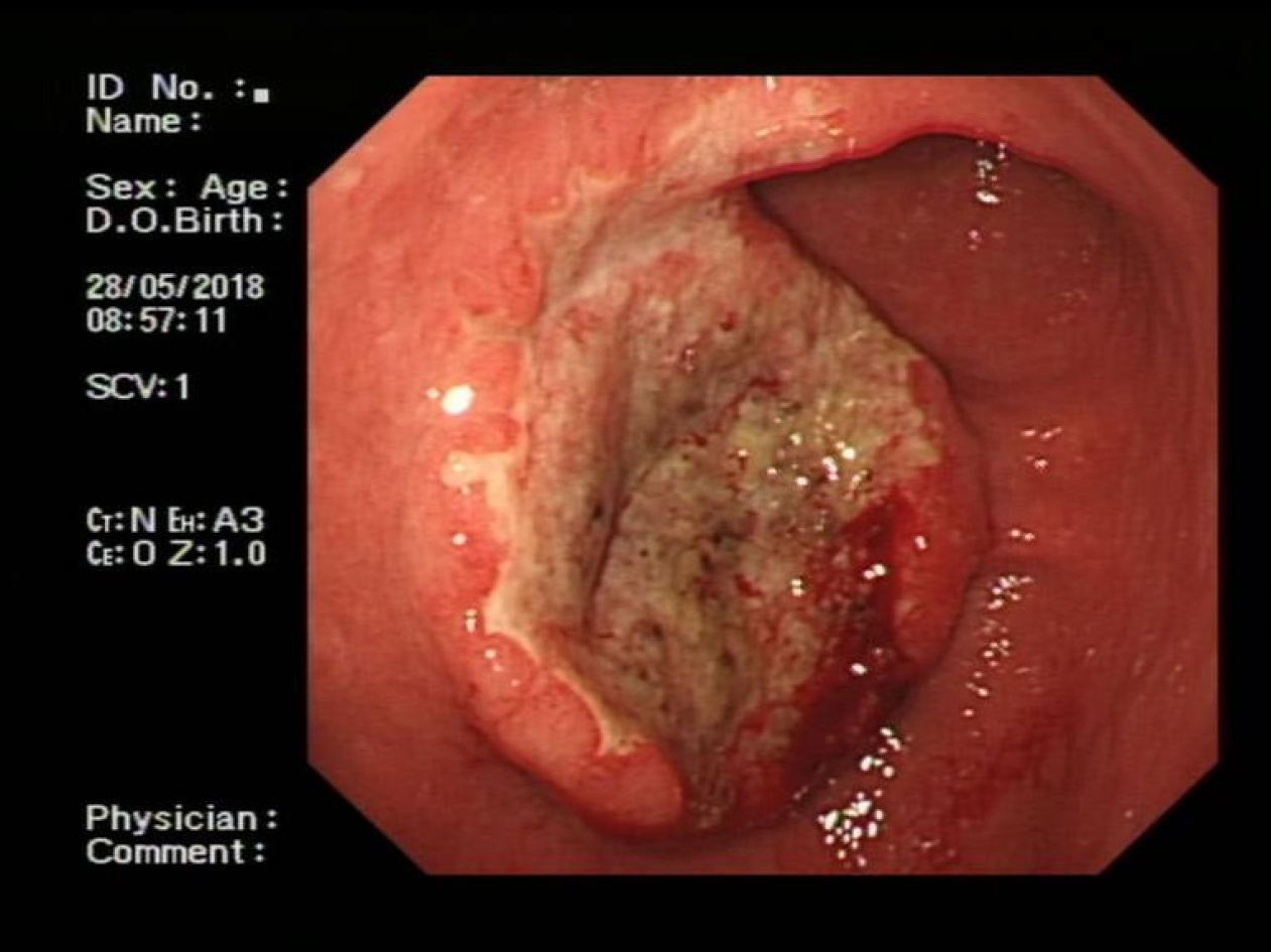
Figure 1 Endoscopic findings of the patient.
Endoscopy showed a large, irregular, raised ulcer 25 mm × 25 mm in dimension at the antrum-body junction.
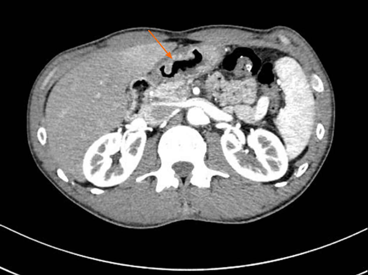
Figure 2 Findings from preoperative abdominal computed tomography.
Abdominal contrast-enhanced computed tomography revealed irregular thickening and enhancement of the wall (arrow).
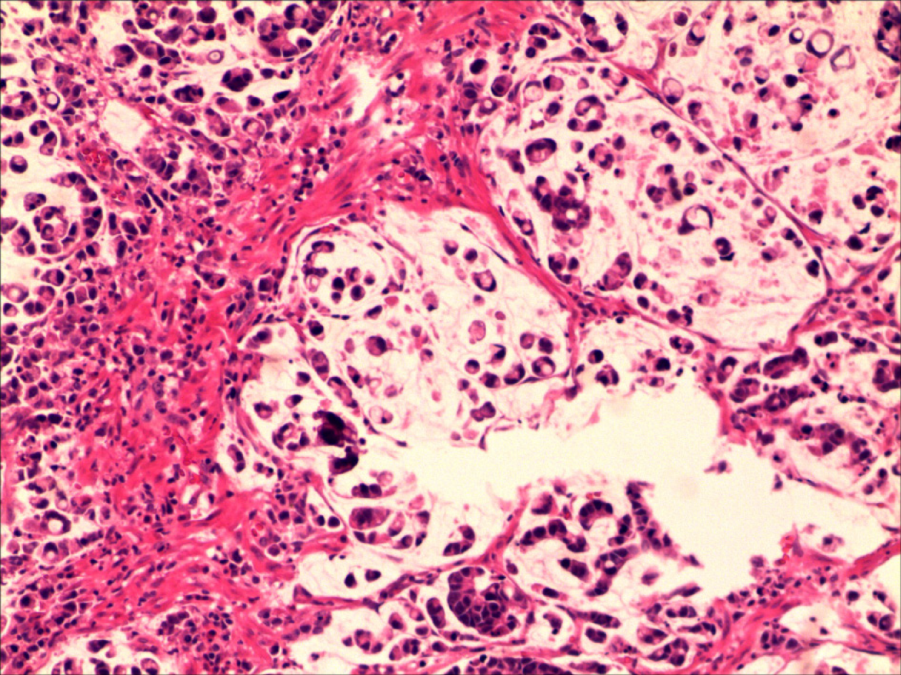
Figure 3 Histological characteristics.
Hematoxylin and eosin stain indicated poorly differentiated adenocarcinoma with signet ring cells (200 × magnification).
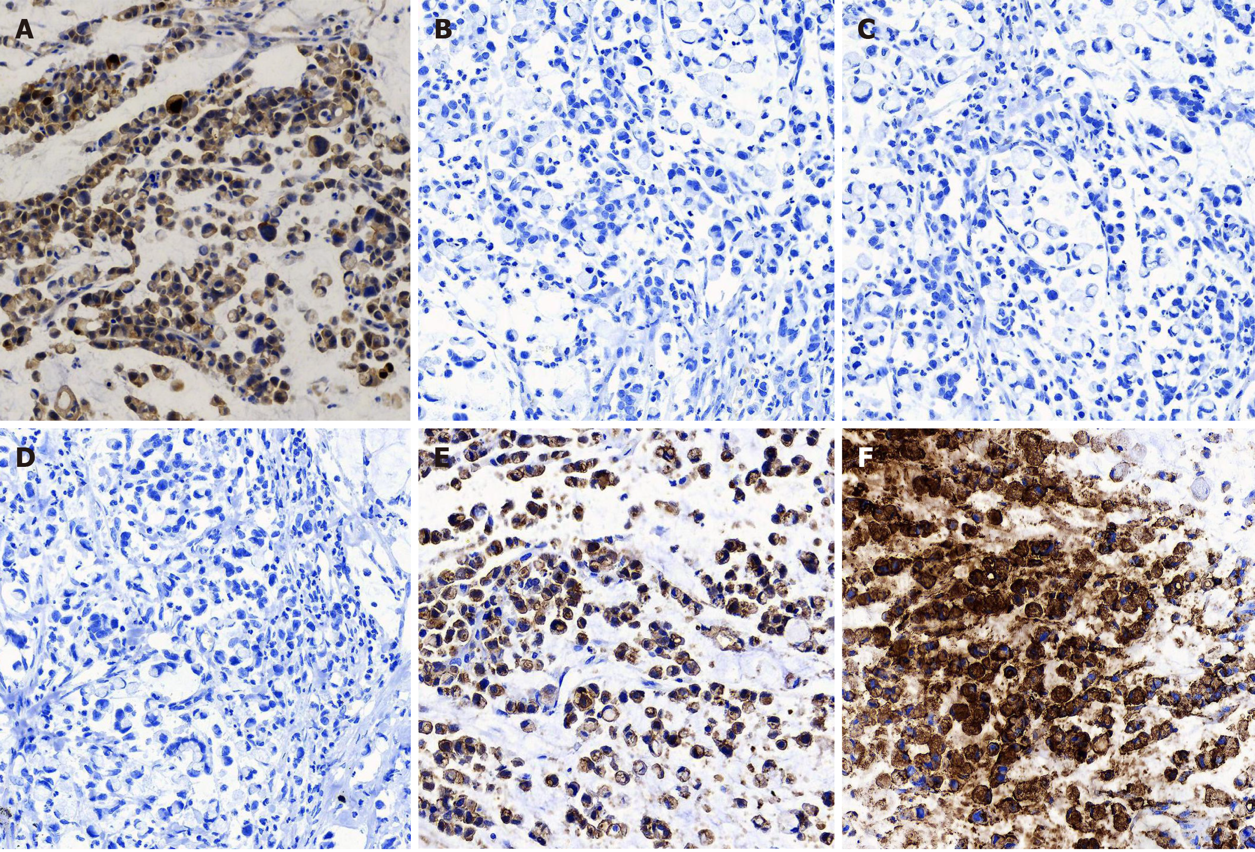
Figure 4 Immunohistochemical staining characteristics.
A: Immunohistochemical (IHC) staining showed strong squamous cell carcinoma antigen expression (+++) mainly in the cytoplasm of tumor cells (200 × magnification); B-D: IHC staining showed that the tumor cells were negative for p40, p63, and CK5/6 (200 × magnification); E and F: IHC staining showed that the tumor cells were positive for E-cadherin and carcinoembryonic antigen (200 × magnification).
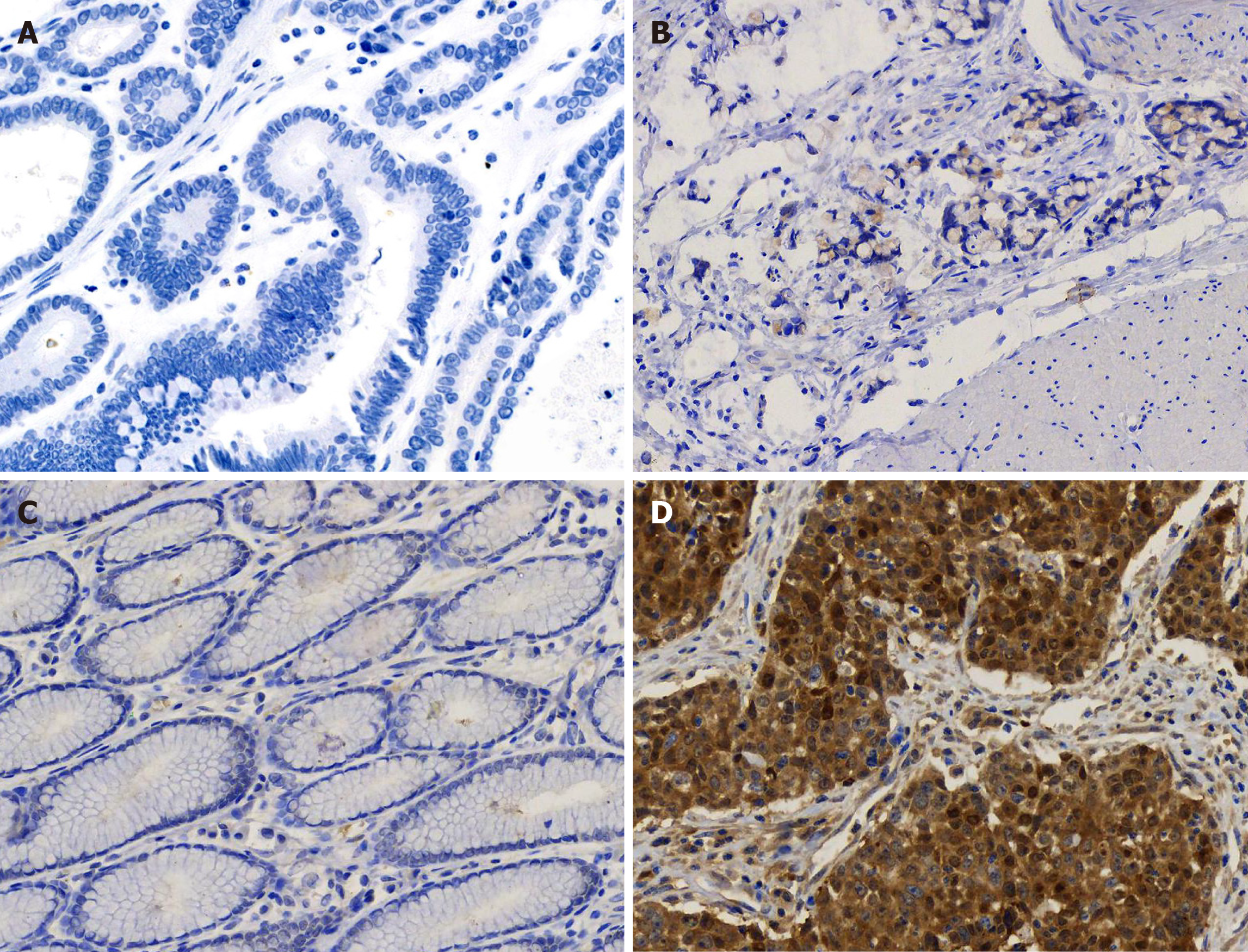
Figure 5 Immunohistochemical staining for squamous cell carcinoma antigen in gastritis and gastric cancer tissues.
Paraffin-sections from gastritis and gastric cancer tissues of multiple patients were simultaneously stained for squamous cell carcinoma antigen (SCCA). A: Negative SCCA expression (-) in well differentiated gastric cancer (200 × magnification); B: Weakly positive SCCA expression (+) in poorly differentiated gastric cancer (200 × magnification); C: Negative SCCA expression (-) in chronic gastritis (200 × magnification); D: Strong SCCA expression (+++) in gastric squamous cell carcinoma (200 × magnification).
- Citation: Wang L, Huang L, Xi L, Zhang SC, Zhang JX. High expression of squamous cell carcinoma antigen in poorly differentiated adenocarcinoma of the stomach: A case report. World J Clin Cases 2020; 8(19): 4572-4578
- URL: https://www.wjgnet.com/2307-8960/full/v8/i19/4572.htm
- DOI: https://dx.doi.org/10.12998/wjcc.v8.i19.4572


