Copyright
©The Author(s) 2020.
World J Clin Cases. Oct 6, 2020; 8(19): 4450-4465
Published online Oct 6, 2020. doi: 10.12998/wjcc.v8.i19.4450
Published online Oct 6, 2020. doi: 10.12998/wjcc.v8.i19.4450
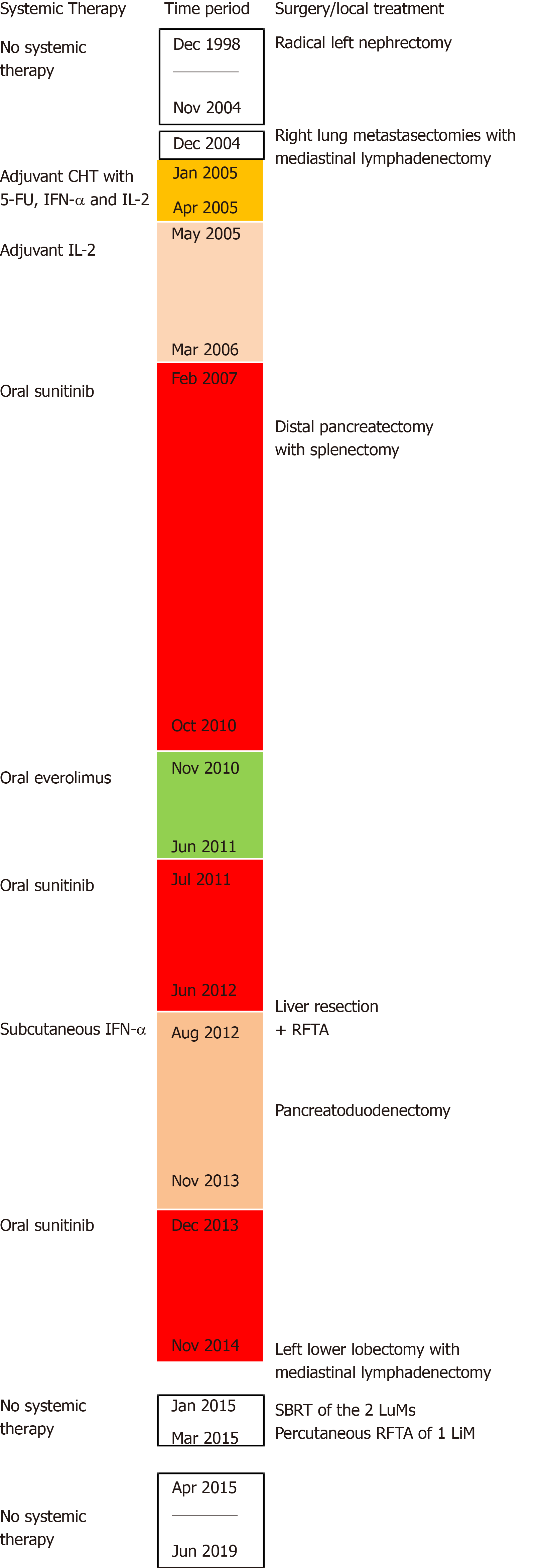
Figure 1 Timeline of systemic therapies, surgical procedures and local treatments.
CHT: Chemotherapy; 5-FU: Fluorouracil; IFN-α: Interferon-alpha; IL-2: Interleukin 2; RFTA: Radiofrequency thermal ablation; SBRT: Stereotactic body radiotherapy; LuM: Lung metastasis; LiM: Liver metastases.
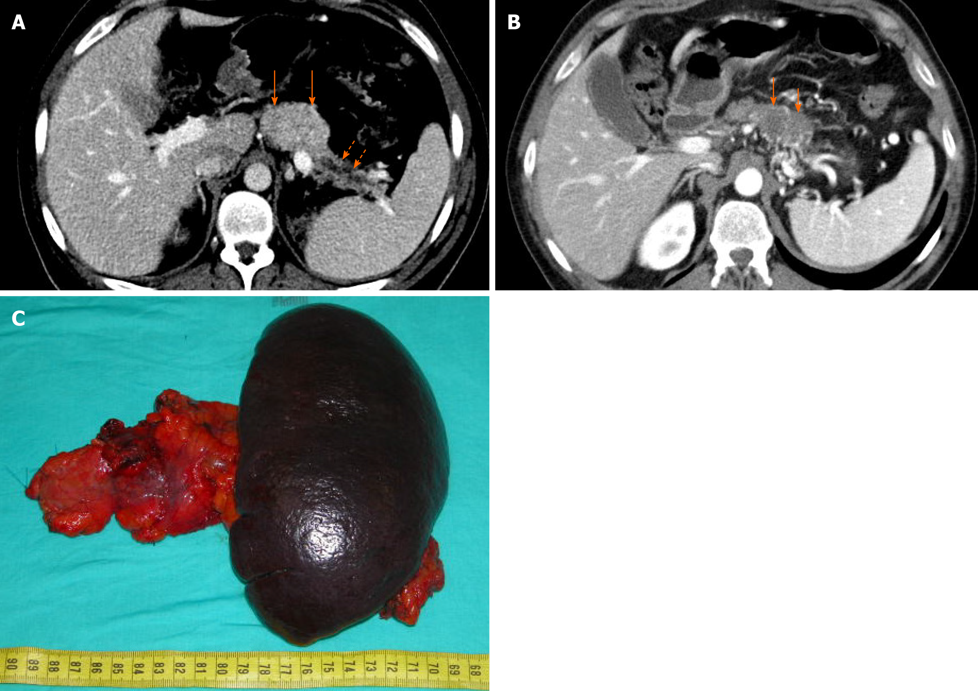
Figure 2 Treatment and outcome of pancreas recurrence.
The contrast-enhanced computerized tomography (CECT) shows a pancreatic metastasis (PaM) measuring 55 mm × 35 mm (A) in the body of the pancreas (orange full arrows), with upstream main pancreatic duct dilatation (orange dotted arrows) and parenchymal atrophy. The CECT performed 5 mo later, after systemic therapy with oral sunitinib, demonstrates a significant volumetric reduction of the PaM (B), which shrunk to 41 mm × 27 mm (orange full arrows). The patient underwent distal pancreatectomy with splenectomy (C).
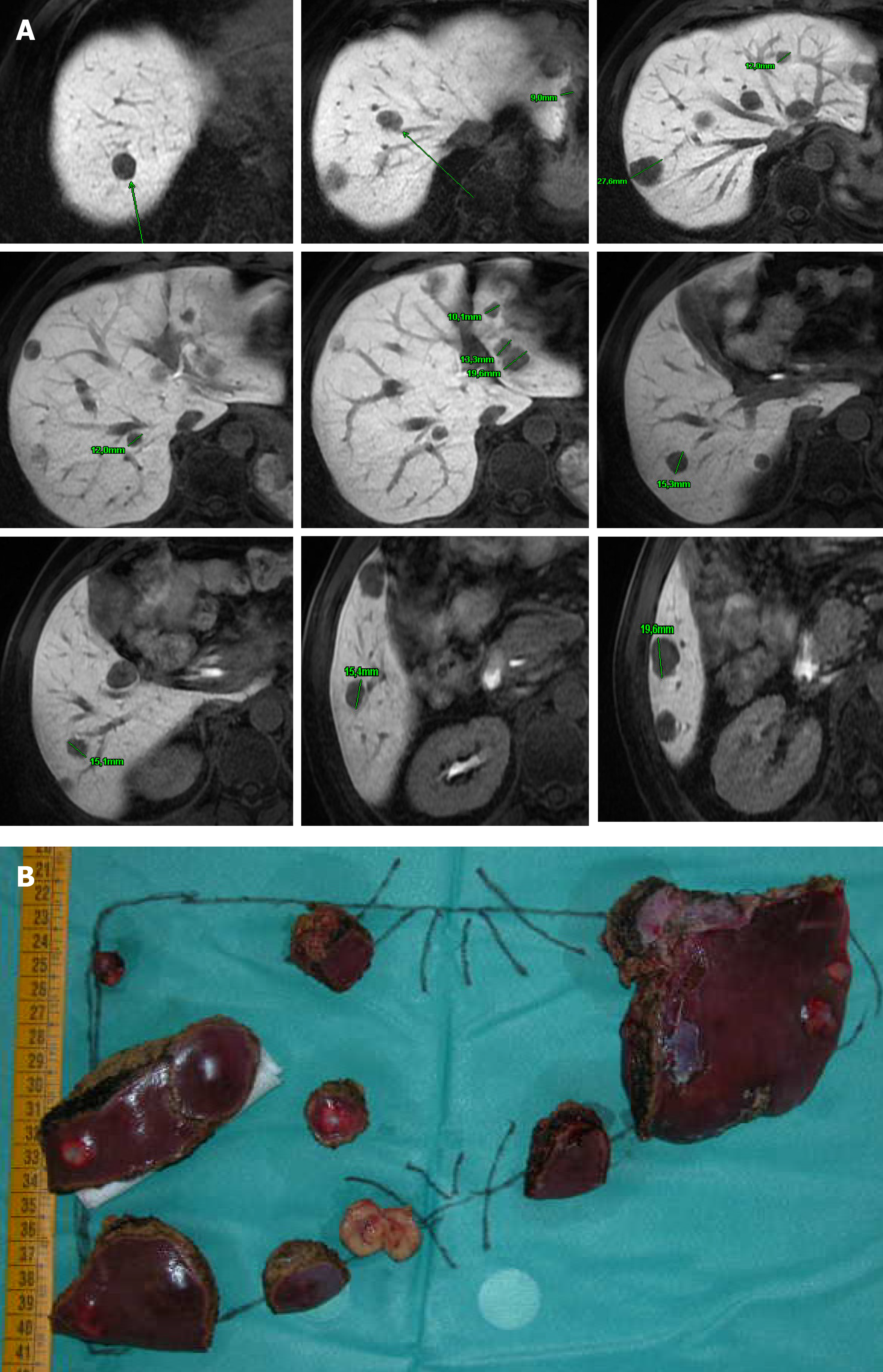
Figure 3 Treament and outcome of liver recurrence.
The magnetic resonance imaging with gadolinium ethoxybenzyl dimeglumine demonstrates multiple liver metastases involving all liver segments except segment S1 (A). The patient underwent intraoperative ultrasonography-guided conservative liver resection (LR) (B), with intermittent hepatic pedicle clamping of 87 min. LR consisted of left lobectomy extended to segment 4a, with gentle detachment of a metastatic nodule from the left portal vein and the middle hepatic vein, respectively; multiple wedge resections in all the remnant liver segments, except S1; and radiofrequency thermal ablation of 3 nodules deeply located in segments S7, S8 and S1-S8, respectively.
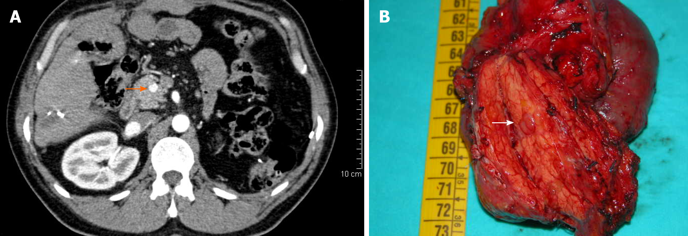
Figure 4 Treatment and outcome of further pancreas recurrence.
The contrast-enhanced computerized tomography scan shows a pancreatic metastasis (PaM) measuring 10 mm in the head of the pancreas (orange full arrow) (A). The patient underwent pancreatoduodenectomy (B); the PaM is indicated in the specimen (white full arrow). Pathological examination revealed multiple intrapancreatic nodules of metastatic clear-cell renal cell carcinoma, without nodal involvement, and with negative surgical margins.
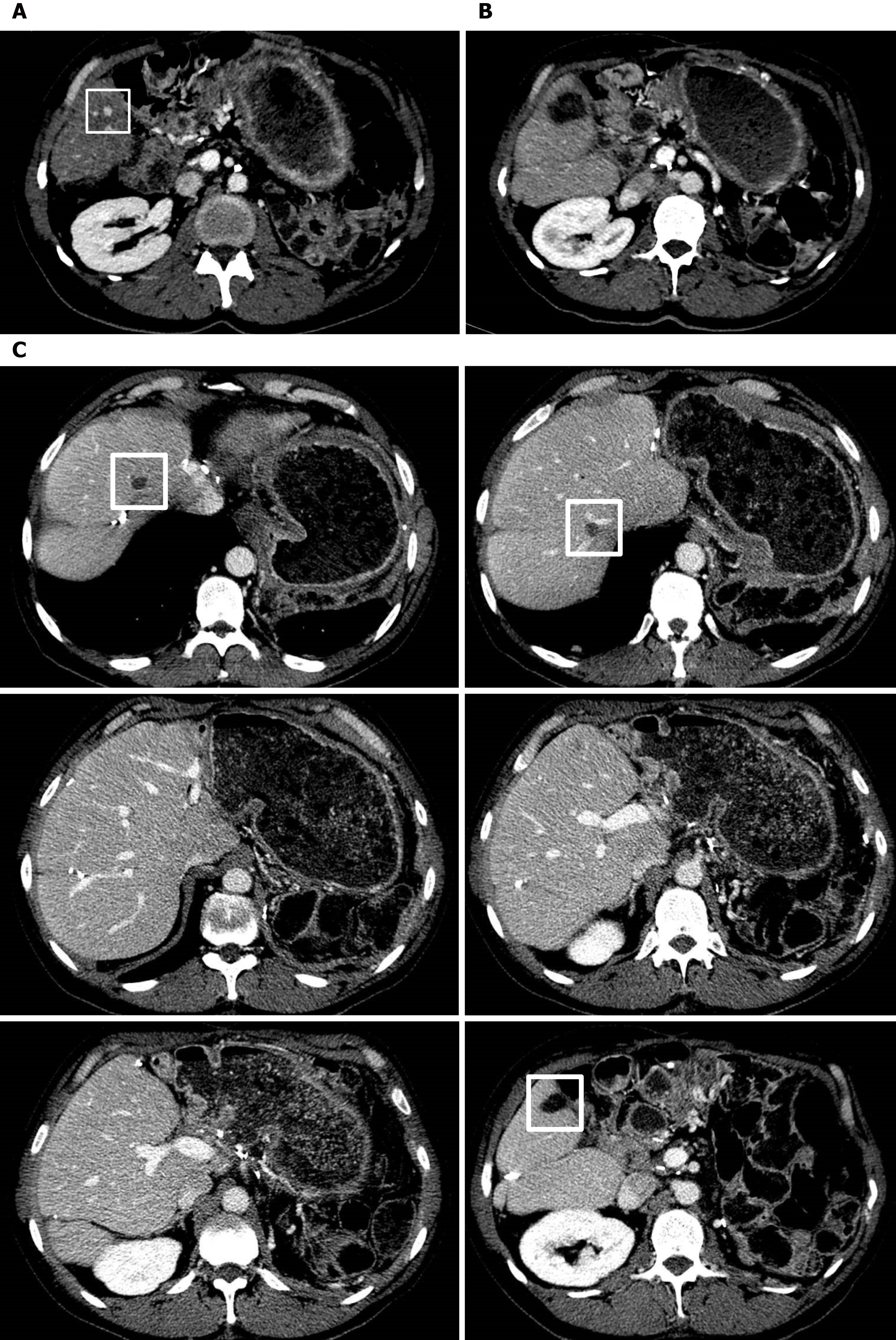
Figure 5 Treatment and outcome of further liver recurrence.
The contrast-enhanced computerized tomography (CECT) scan demonstrates a liver metastases (LiM) measuring 8 mm in segment S6 (white square) (A). The patient underwent successful percutaneous radiofrequency thermal ablation (RFTA), as shown in the postoperative CECT (B). The CECT scan 18 mo later demonstrates the absence of further LiMs, with the areas of intraoperative RFTA in segments S8 and S7, respectively, and the area of percutaneous RFTA in segment S6, delineated by white squares (C).
- Citation: De Raffele E, Mirarchi M, Casadei R, Ricci C, Brunocilla E, Minni F. Twenty-year survival after iterative surgery for metastatic renal cell carcinoma: A case report and review of literature. World J Clin Cases 2020; 8(19): 4450-4465
- URL: https://www.wjgnet.com/2307-8960/full/v8/i19/4450.htm
- DOI: https://dx.doi.org/10.12998/wjcc.v8.i19.4450


