Copyright
©The Author(s) 2020.
World J Clin Cases. Oct 6, 2020; 8(19): 4450-4465
Published online Oct 6, 2020. doi: 10.12998/wjcc.v8.i19.4450
Published online Oct 6, 2020. doi: 10.12998/wjcc.v8.i19.4450
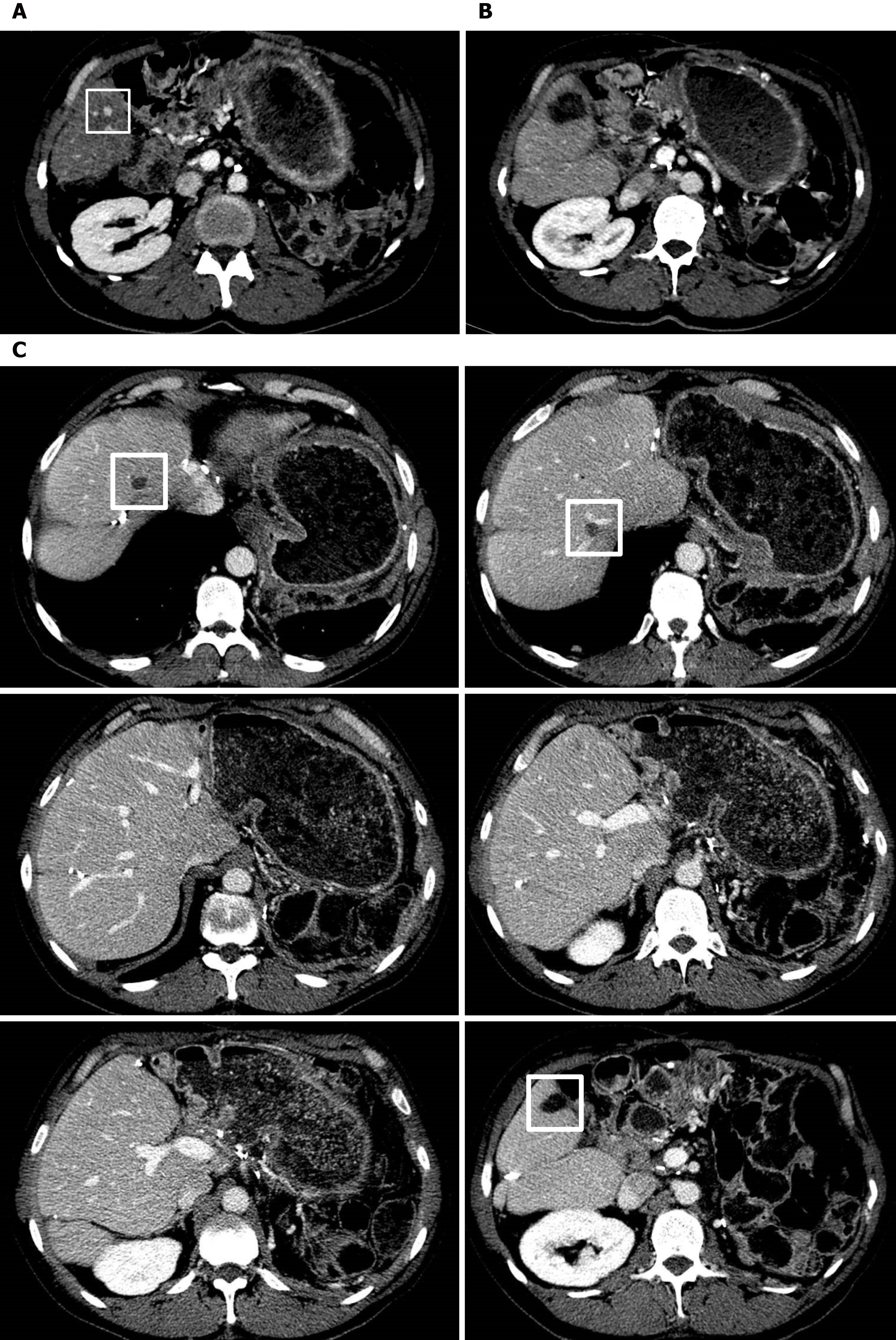
Figure 5 Treatment and outcome of further liver recurrence.
The contrast-enhanced computerized tomography (CECT) scan demonstrates a liver metastases (LiM) measuring 8 mm in segment S6 (white square) (A). The patient underwent successful percutaneous radiofrequency thermal ablation (RFTA), as shown in the postoperative CECT (B). The CECT scan 18 mo later demonstrates the absence of further LiMs, with the areas of intraoperative RFTA in segments S8 and S7, respectively, and the area of percutaneous RFTA in segment S6, delineated by white squares (C).
- Citation: De Raffele E, Mirarchi M, Casadei R, Ricci C, Brunocilla E, Minni F. Twenty-year survival after iterative surgery for metastatic renal cell carcinoma: A case report and review of literature. World J Clin Cases 2020; 8(19): 4450-4465
- URL: https://www.wjgnet.com/2307-8960/full/v8/i19/4450.htm
- DOI: https://dx.doi.org/10.12998/wjcc.v8.i19.4450


