Copyright
©The Author(s) 2020.
World J Clin Cases. Aug 6, 2020; 8(15): 3284-3290
Published online Aug 6, 2020. doi: 10.12998/wjcc.v8.i15.3284
Published online Aug 6, 2020. doi: 10.12998/wjcc.v8.i15.3284
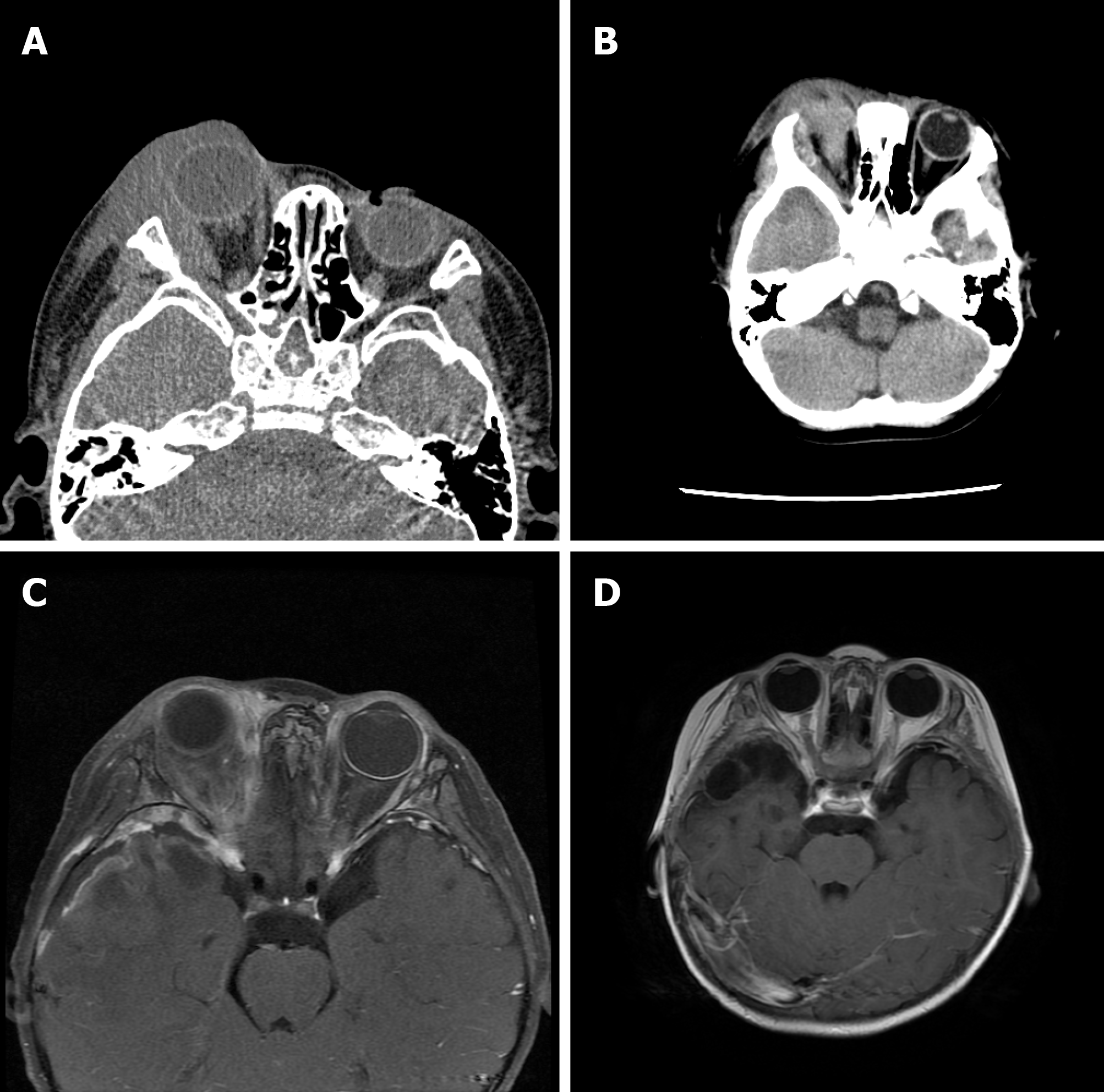
Figure 1 Imaging examinations.
A: Eyelid computed tomography (CT) at admission. A soft tissue mass in the right lateral eyelid muscle was noted; B: CT scan of the head after surgery. The lesion range had increased, involving the temporal lobe; C: Enhanced magnetic resonance imaging (MRI) of the eyelid after the first chemotherapy. The right eyelid and its surrounding space-occupying lesion as well as abnormal signal in the right temporal lobe were visible; D: Brain enhanced MRI after treatment. The original lesion was significantly reduced and disappeared.
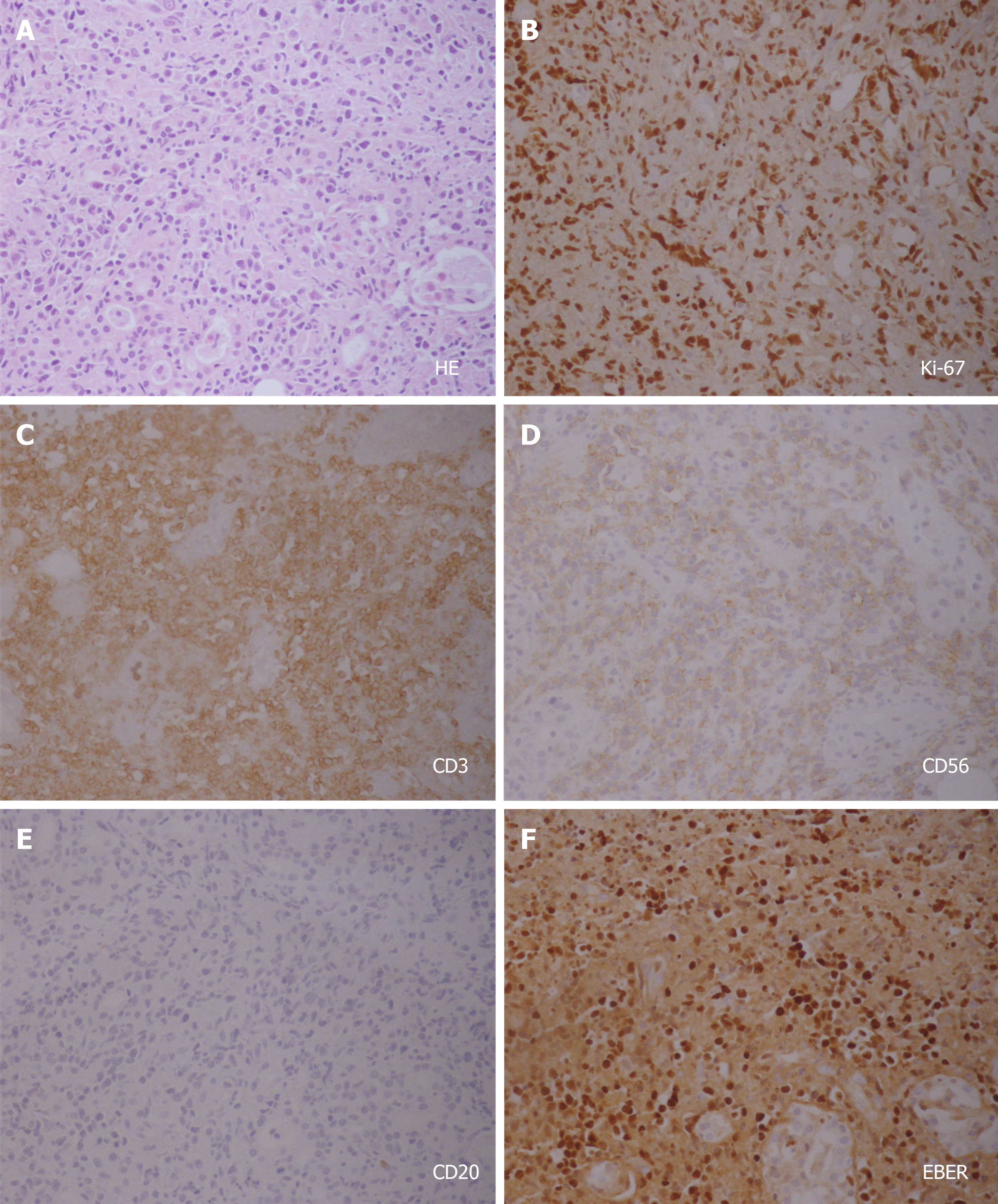
Figure 2 Pathological outcomes.
A: Hematoxylin and eosin staining showing necrosis in the tissue, and atypical lymphocytes infiltrated between the glands; B-F: Immunohistochemical staining showing (B) a Ki-67 index of 80%, (C) CD3+, (D) CD56+, (E) CD20-, and (F) EBER+. Original magnification for all images: 10 × 20. HE: Hematoxylin and eosin.
- Citation: Li N, Wang YZ, Zhang Y, Zhang WL, Zhou Y, Huang DS. Natural killer/T-cell lymphoma with intracranial infiltration and Epstein-Barr virus infection: A case report. World J Clin Cases 2020; 8(15): 3284-3290
- URL: https://www.wjgnet.com/2307-8960/full/v8/i15/3284.htm
- DOI: https://dx.doi.org/10.12998/wjcc.v8.i15.3284


