Copyright
©The Author(s) 2019.
World J Clin Cases. Aug 6, 2019; 7(15): 2058-2064
Published online Aug 6, 2019. doi: 10.12998/wjcc.v7.i15.2058
Published online Aug 6, 2019. doi: 10.12998/wjcc.v7.i15.2058
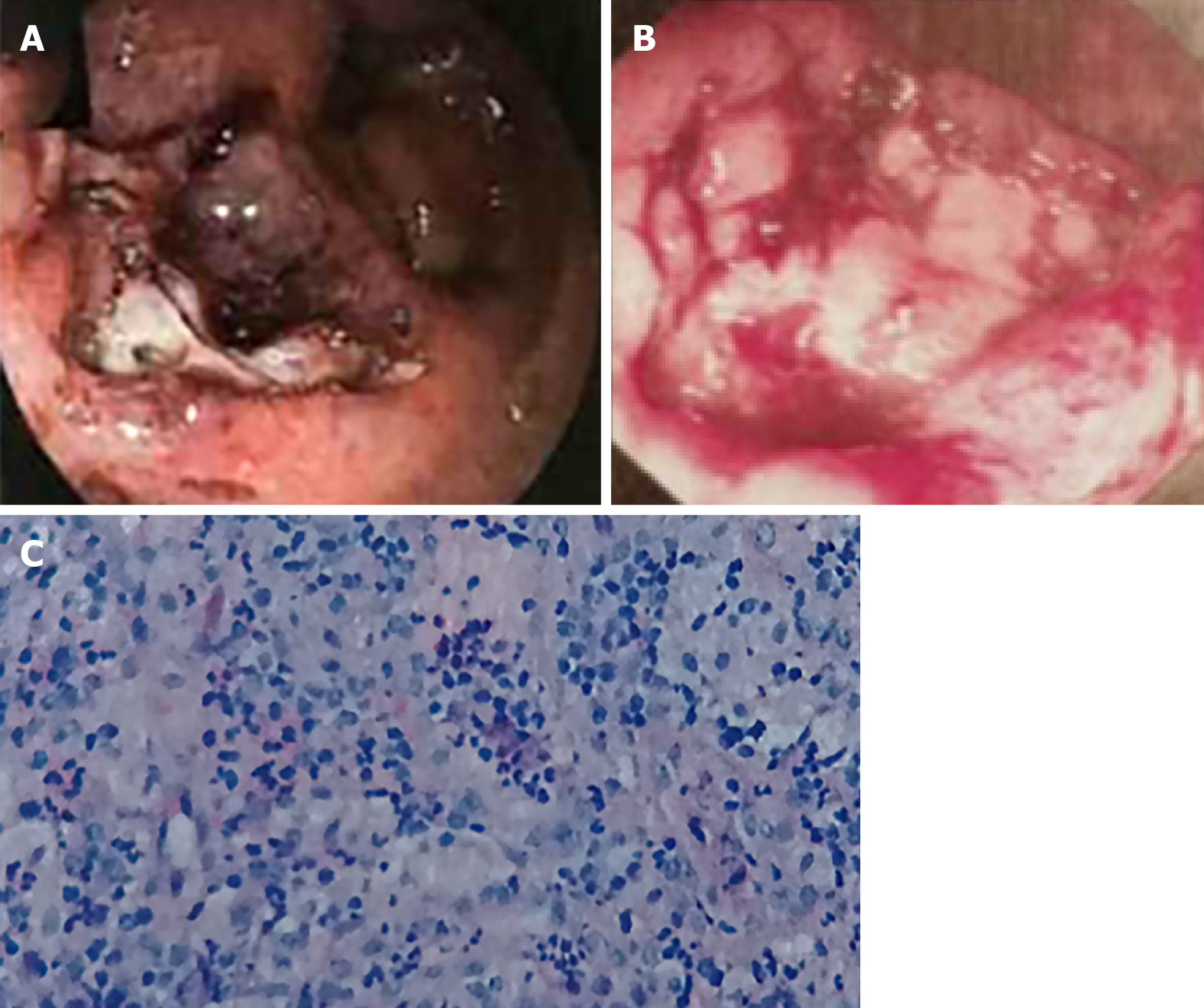
Figure 1 Endoscopic images and corresponding histological findings from before the Traditional Chinese Medicine treatment.
A: Colonoscopy image showing actively bleeding, deep ulcerations; B: Rectoscope image showing a large ulcerative lesion covered by a white and bloody sloughing tissue; C: Hematoxylin-eosin-stained rectal ulcers.
Figure 2 Endoscopic imaging and corresponding histological findings after Traditional Chinese Medicine treatment.
A: The improvement of ulcers; B: Hematoxylin-eosin-stained rectal ulcers (× 40); C: The healed ulcers.
- Citation: Zhang LL, Hao WS, Xu M, Li C, Shi YY. Modified Tong Xie Yao Fang relieves solitary rectal ulcer syndrome: A case report. World J Clin Cases 2019; 7(15): 2058-2064
- URL: https://www.wjgnet.com/2307-8960/full/v7/i15/2058.htm
- DOI: https://dx.doi.org/10.12998/wjcc.v7.i15.2058


