Copyright
©The Author(s) 2019.
World J Clin Cases. Jul 6, 2019; 7(13): 1582-1590
Published online Jul 6, 2019. doi: 10.12998/wjcc.v7.i13.1582
Published online Jul 6, 2019. doi: 10.12998/wjcc.v7.i13.1582
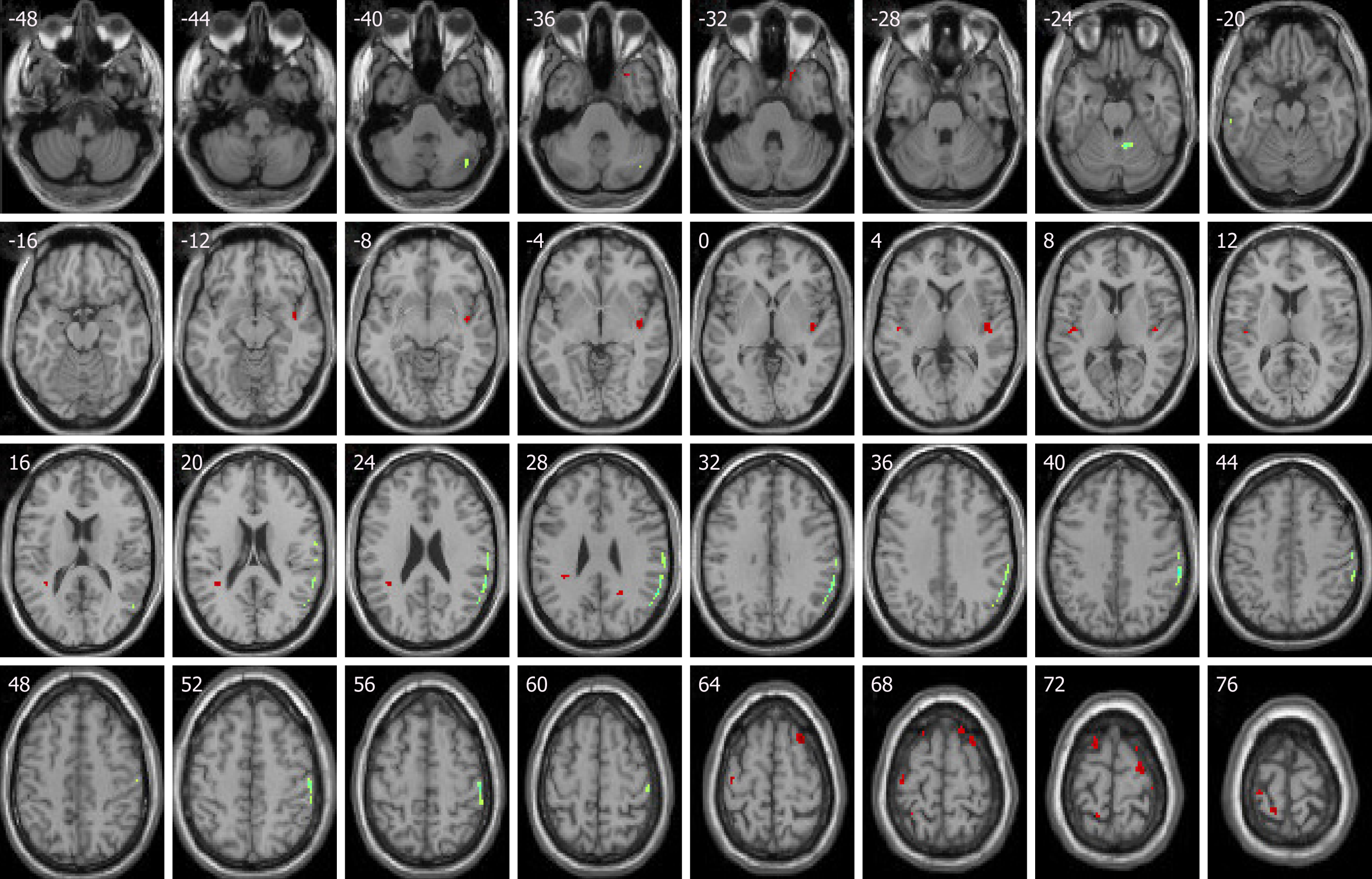
Figure 1 Compared with baseline levels, idiopathic tinnitus patients before treatment showed increased activities in the right parahippocampa gyrus, right superior temporal gyrus, right superior frontal gyrus, anterior insula, left inferior parietal lobule, and left precentral gyrus.
Decreased activities were in left postcentral gyrus and left ITG. (Red shows increased activity, and green shows decreased activity.)
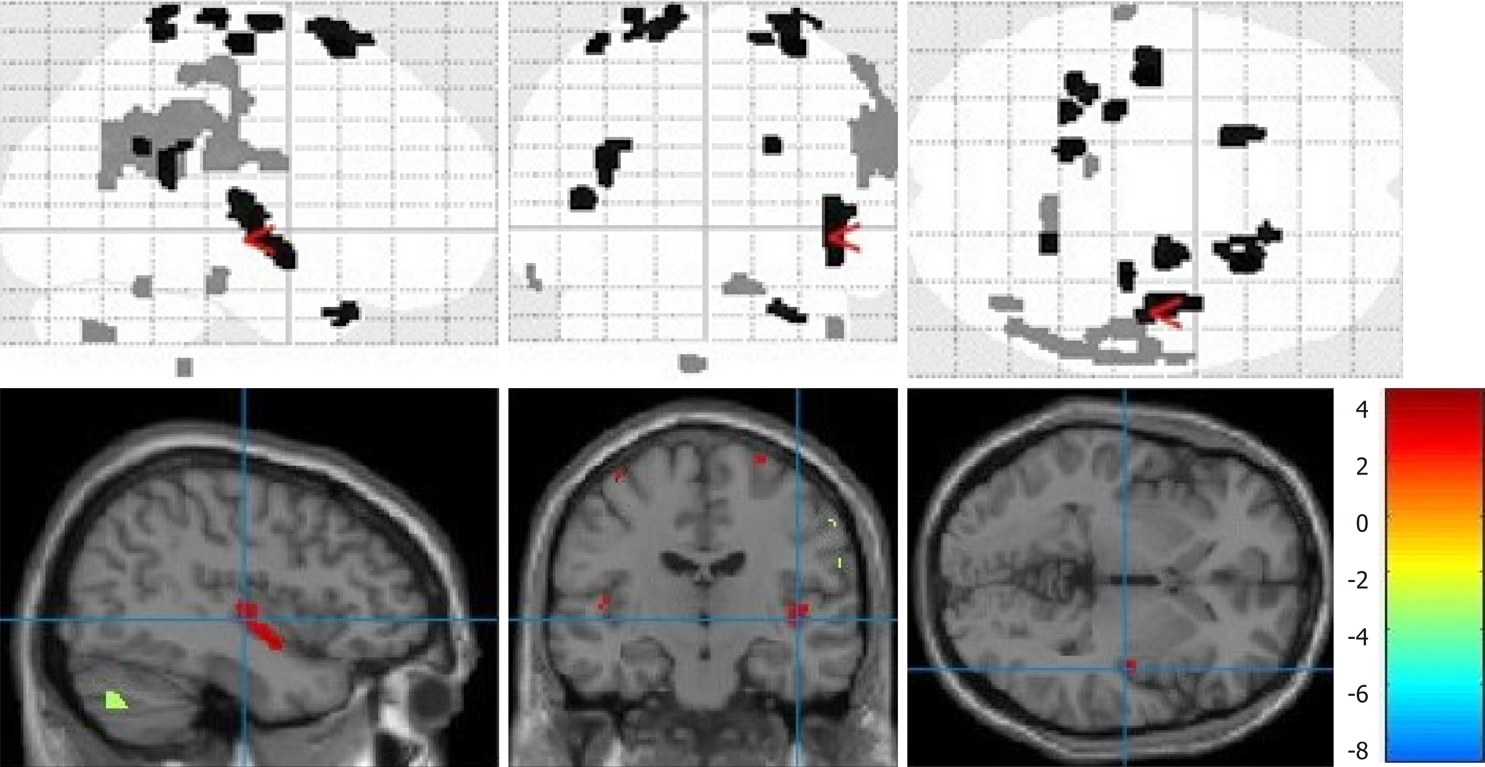
Figure 2 Volume render and matrix of statistical parametric mapping.
- Citation: Kan Y, Wang W, Zhang SX, Ma H, Wang ZC, Yang JG. Neural metabolic activity in idiopathic tinnitus patients after repetitive transcranial magnetic stimulation. World J Clin Cases 2019; 7(13): 1582-1590
- URL: https://www.wjgnet.com/2307-8960/full/v7/i13/1582.htm
- DOI: https://dx.doi.org/10.12998/wjcc.v7.i13.1582


