Copyright
©The Author(s) 2023.
World J Clin Cases. Feb 6, 2023; 11(4): 896-902
Published online Feb 6, 2023. doi: 10.12998/wjcc.v11.i4.896
Published online Feb 6, 2023. doi: 10.12998/wjcc.v11.i4.896
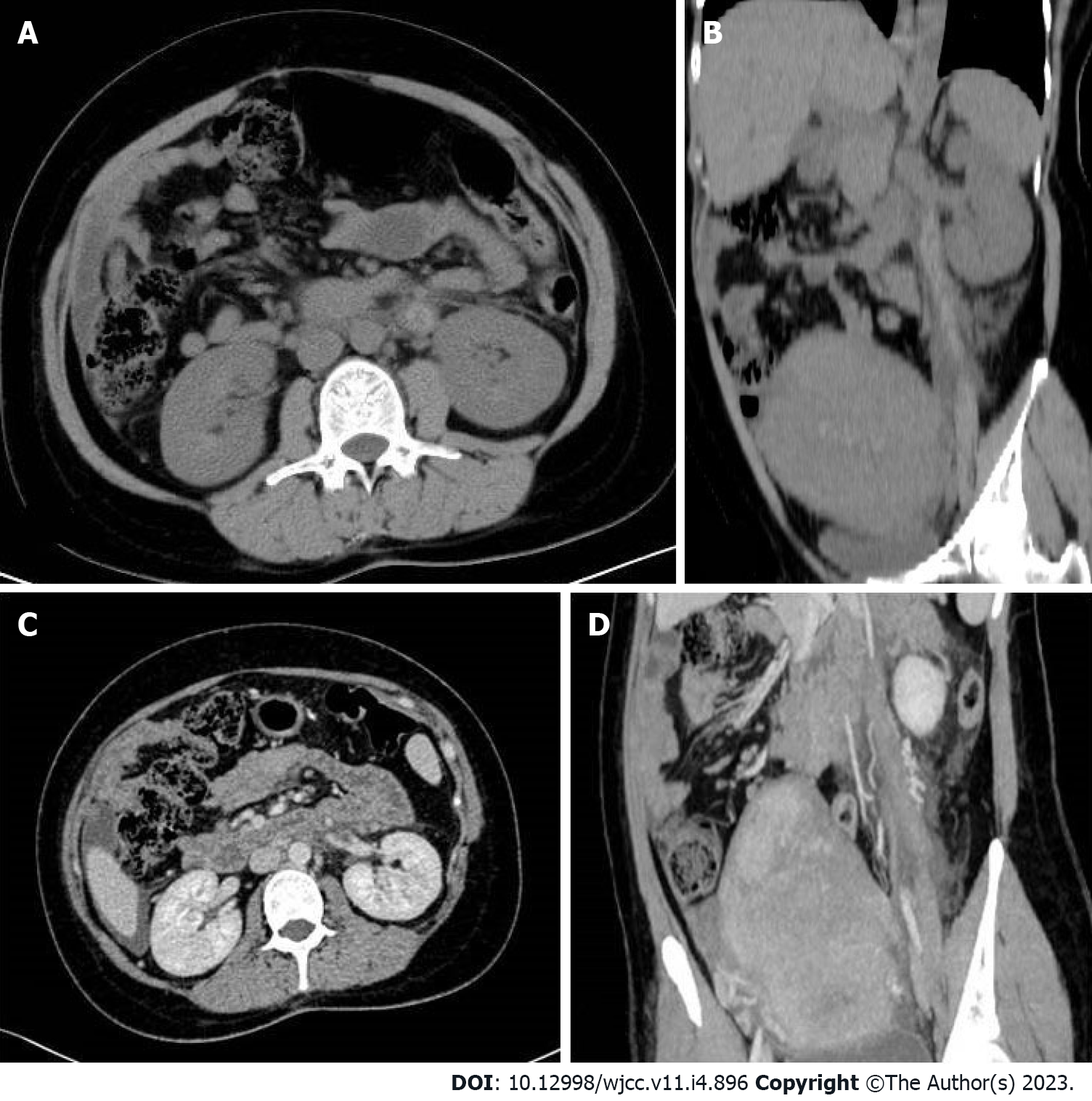
Figure 1 Computed tomography images.
A and B: Plain computed tomography images; C and D: Enhanced computed tomography images.
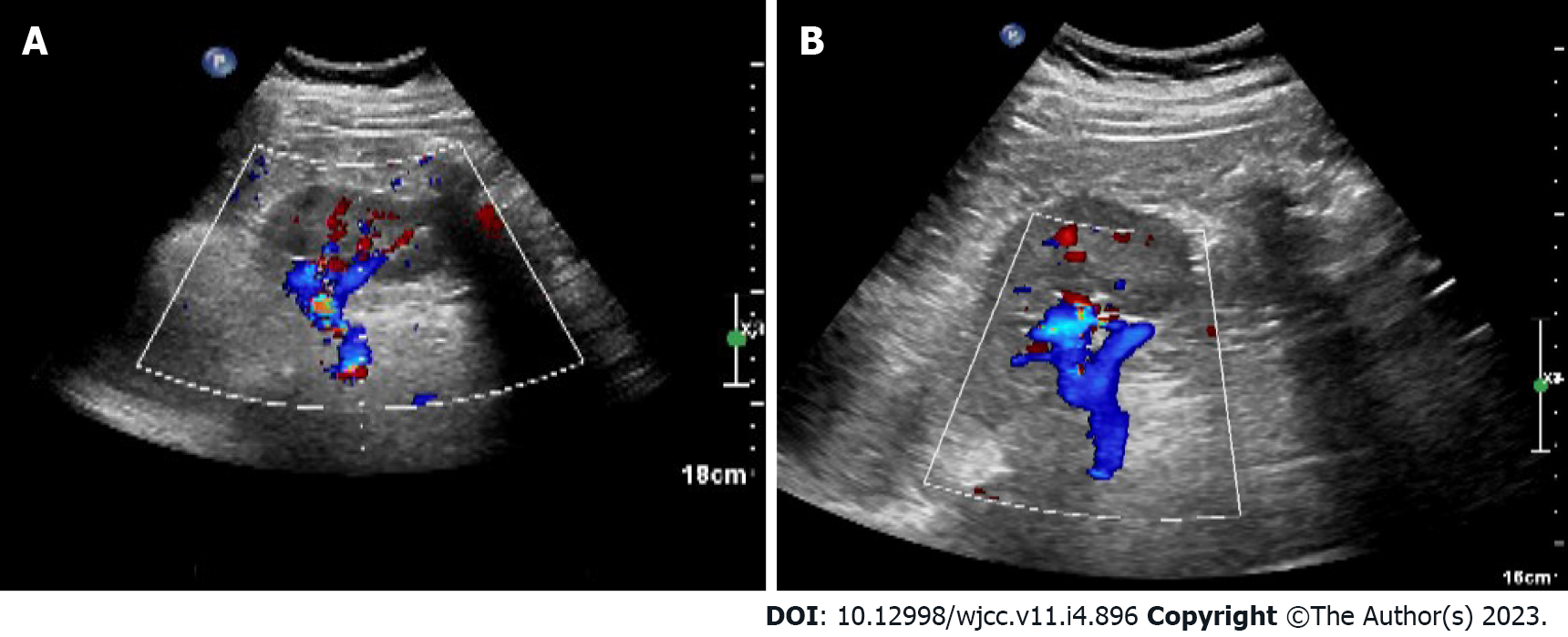
Figure 2 B-ultrasound performed at one month postpartum showed no abnormalities in bilateral ovaries and uterus.
A: Sagittal view 1; B: Sagittal view 2.
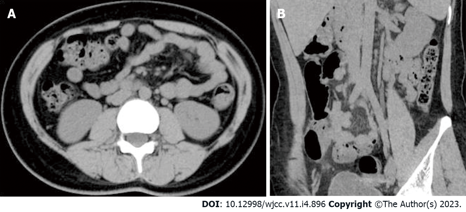
Figure 3 Plain computed tomography images six months postpartum.
A: Coronal view; B: Sagittal view.
- Citation: Wang JJ, Hui CC, Ji YD, Xu W. Computed tomography diagnosed left ovarian venous thrombophlebitis after vaginal delivery: A case report. World J Clin Cases 2023; 11(4): 896-902
- URL: https://www.wjgnet.com/2307-8960/full/v11/i4/896.htm
- DOI: https://dx.doi.org/10.12998/wjcc.v11.i4.896


