Copyright
©The Author(s) 2023.
World J Clin Cases. Feb 6, 2023; 11(4): 852-858
Published online Feb 6, 2023. doi: 10.12998/wjcc.v11.i4.852
Published online Feb 6, 2023. doi: 10.12998/wjcc.v11.i4.852
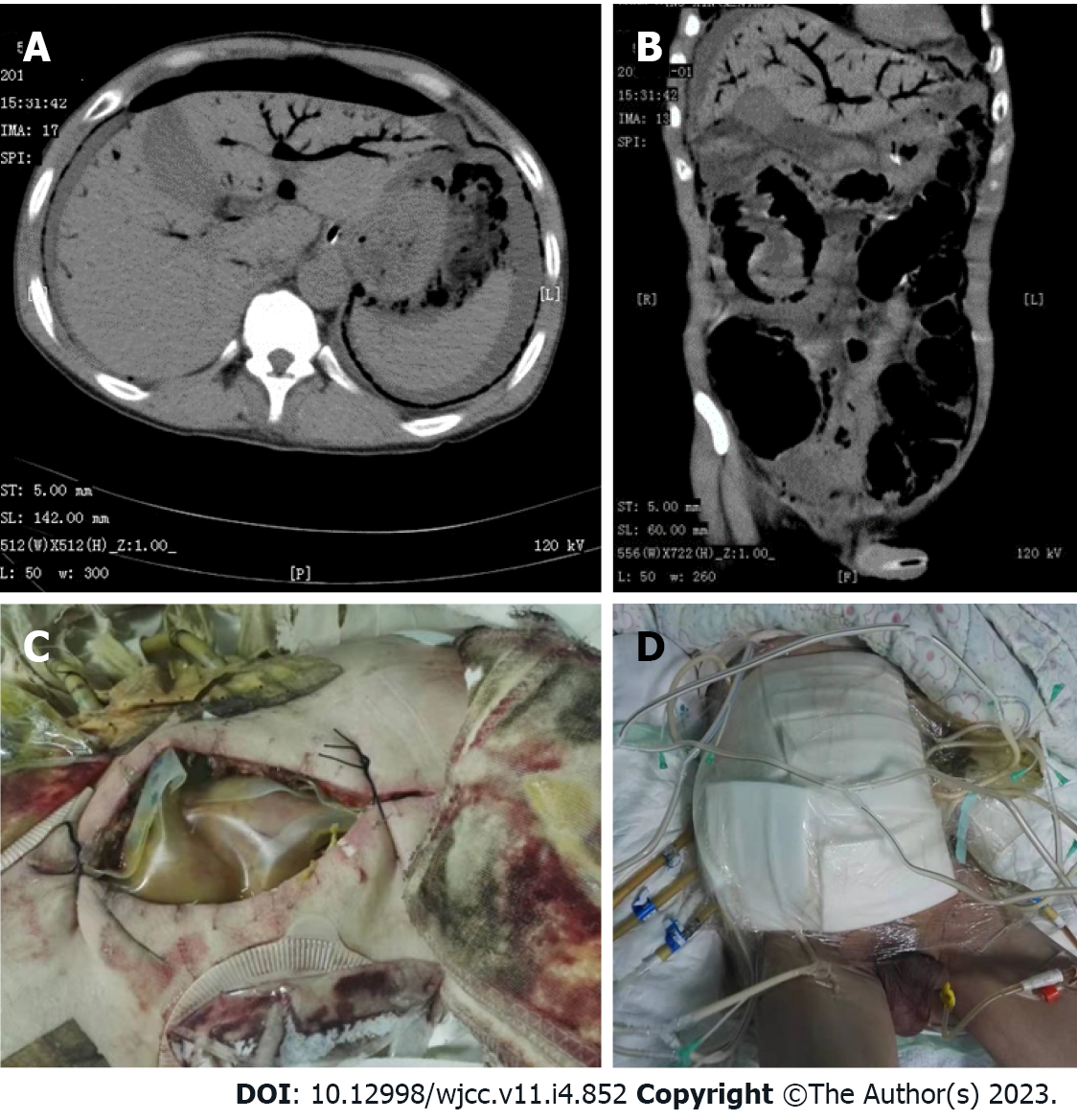
Figure 1 Image examination and treatment.
A: Preoperative computed tomography (CT) showing pneumoperitoneum and portal venous gas; B: Coronal view of abdominal CT showing extensive portal venous gas; C: Bogota bag before the second surgery; D: Vacuum sealing drainage.
- Citation: Li HY, Wang ZX, Wang JC, Zhang XD. Clostridium perfringens gas gangrene caused by closed abdominal injury: A case report and review of the literature. World J Clin Cases 2023; 11(4): 852-858
- URL: https://www.wjgnet.com/2307-8960/full/v11/i4/852.htm
- DOI: https://dx.doi.org/10.12998/wjcc.v11.i4.852


