Copyright
©The Author(s) 2022.
World J Clin Cases. Dec 16, 2022; 10(35): 12971-12979
Published online Dec 16, 2022. doi: 10.12998/wjcc.v10.i35.12971
Published online Dec 16, 2022. doi: 10.12998/wjcc.v10.i35.12971
Figure 1 Computed tomography scan of the abdomen.
A and B: Preoperative computed tomography examination findings showed free intraperitoneal air (A) and intraperitoneal free fluid (B) in case 1; C: Computed tomography scan of the abdomen revealed free intraperitoneal air under the diaphragm and in the right abdominal area in case 2.
Figure 2 Intraoperative findings.
Laparotomy showed patchy necrosis and subserosal white plaque lesions on the small bowel wall, along with multiple perforations. A: Patchy necrosis and subserosal white plaque lesions on the small bowel wall in case 1; B: Multiple perforations and subserosal white plaque lesions in case 1; C: Operative findings during initial exploratory laparotomy showed multiple perforations at the distal ileum in case 2.
Figure 3 Cutaneous lesions with aubergine papules on the lower extremities.
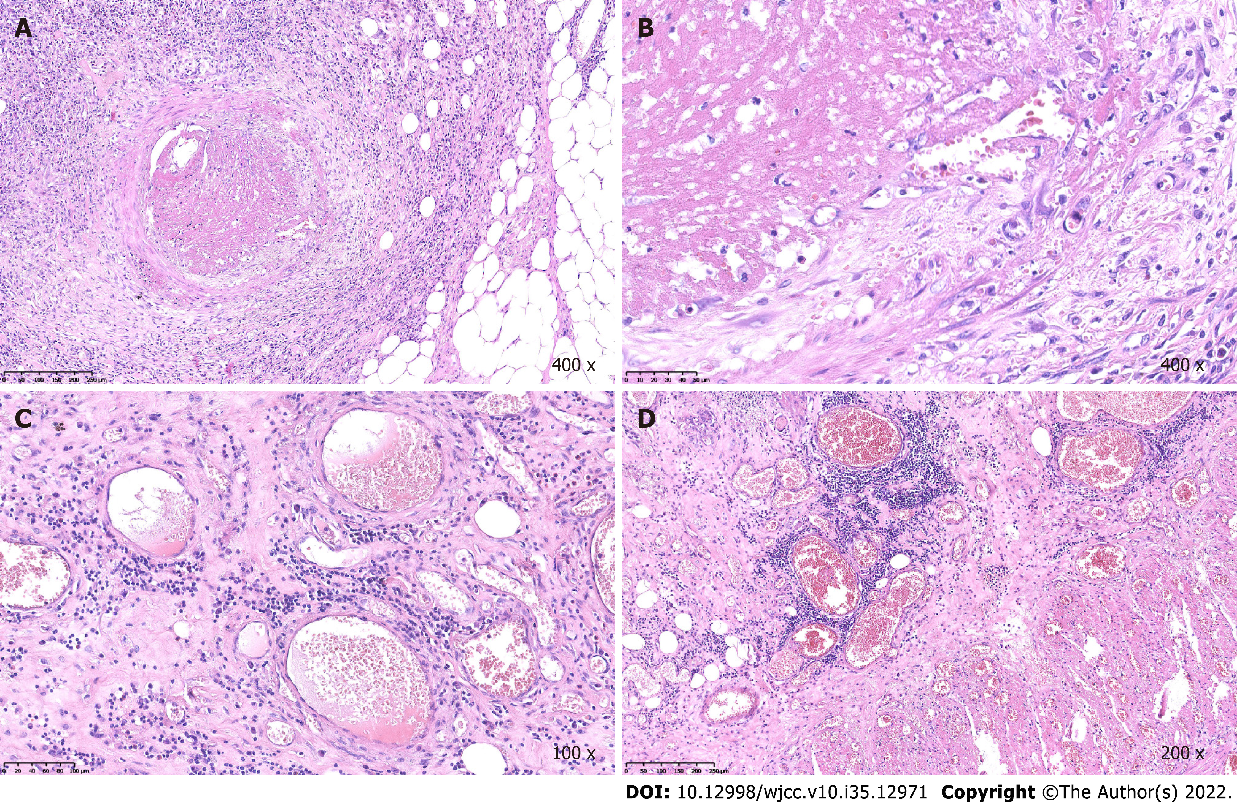
Figure 4 Histological findings of the gastrointestinal tract in case 1.
A and B: Representative histological findings of the small bowel, which revealed extensive hyperemia, edema and necrosis (hematoxylin and eosin staining), as well as small vasculitis with fibrinoid necrosis and intraluminal thrombosis; C and D: Representative histological findings which showed inflammatory cell infiltration and congestion of the intestinal vasculature.
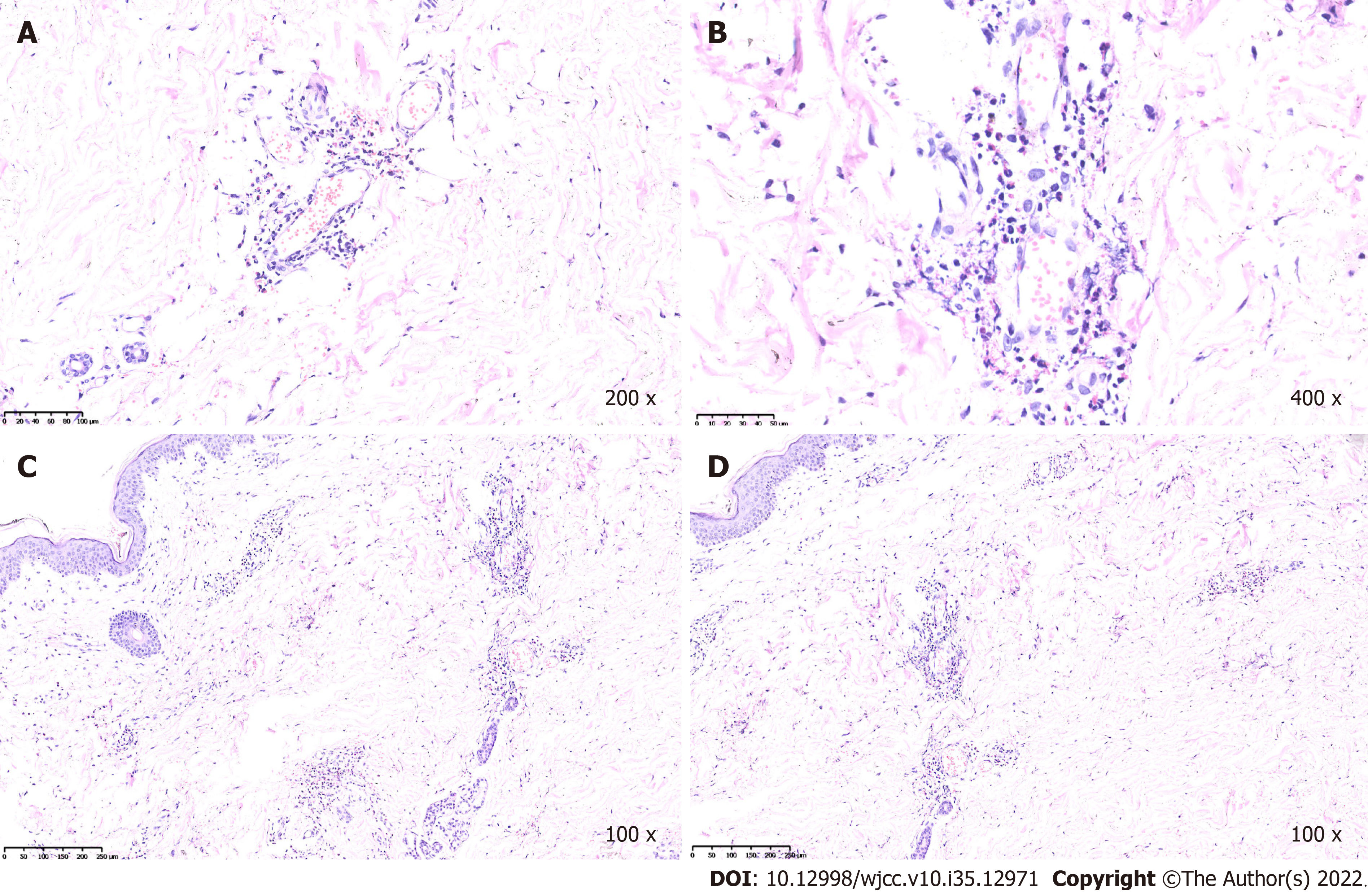
Figure 5 Histological findings of the skin in case 1.
A and B: Representative sections from biopsies of the cutaneous lesions, which revealed the swollen endothelium of small vessels in the dermis, perivascular eosinophils, neutrophils, lymphocytes and broken leukocyte infiltration (hematoxylin and eosin staining); C and D: Representative sections from biopsies, which showed the swollen endothelium of small vessels in the dermis. Perivascular eosinophils, neutrophils, lymphocytes and broken leukocyte infiltration.
Figure 6 Postoperative digestive tract radiography and gastroscopy in case 2.
A: Esophageal perforation was shown in postoperative computed tomography images; B-D: Representative images of gross anatomy showing ulcerations at the fundus and cardia of the stomach and esophagus.
- Citation: Li ZG, Zhou JM, Li L, Wang XD. Malignant atrophic papulosis: Two case reports. World J Clin Cases 2022; 10(35): 12971-12979
- URL: https://www.wjgnet.com/2307-8960/full/v10/i35/12971.htm
- DOI: https://dx.doi.org/10.12998/wjcc.v10.i35.12971


