Copyright
©The Author(s) 2022.
World J Clin Cases. Dec 16, 2022; 10(35): 12971-12979
Published online Dec 16, 2022. doi: 10.12998/wjcc.v10.i35.12971
Published online Dec 16, 2022. doi: 10.12998/wjcc.v10.i35.12971
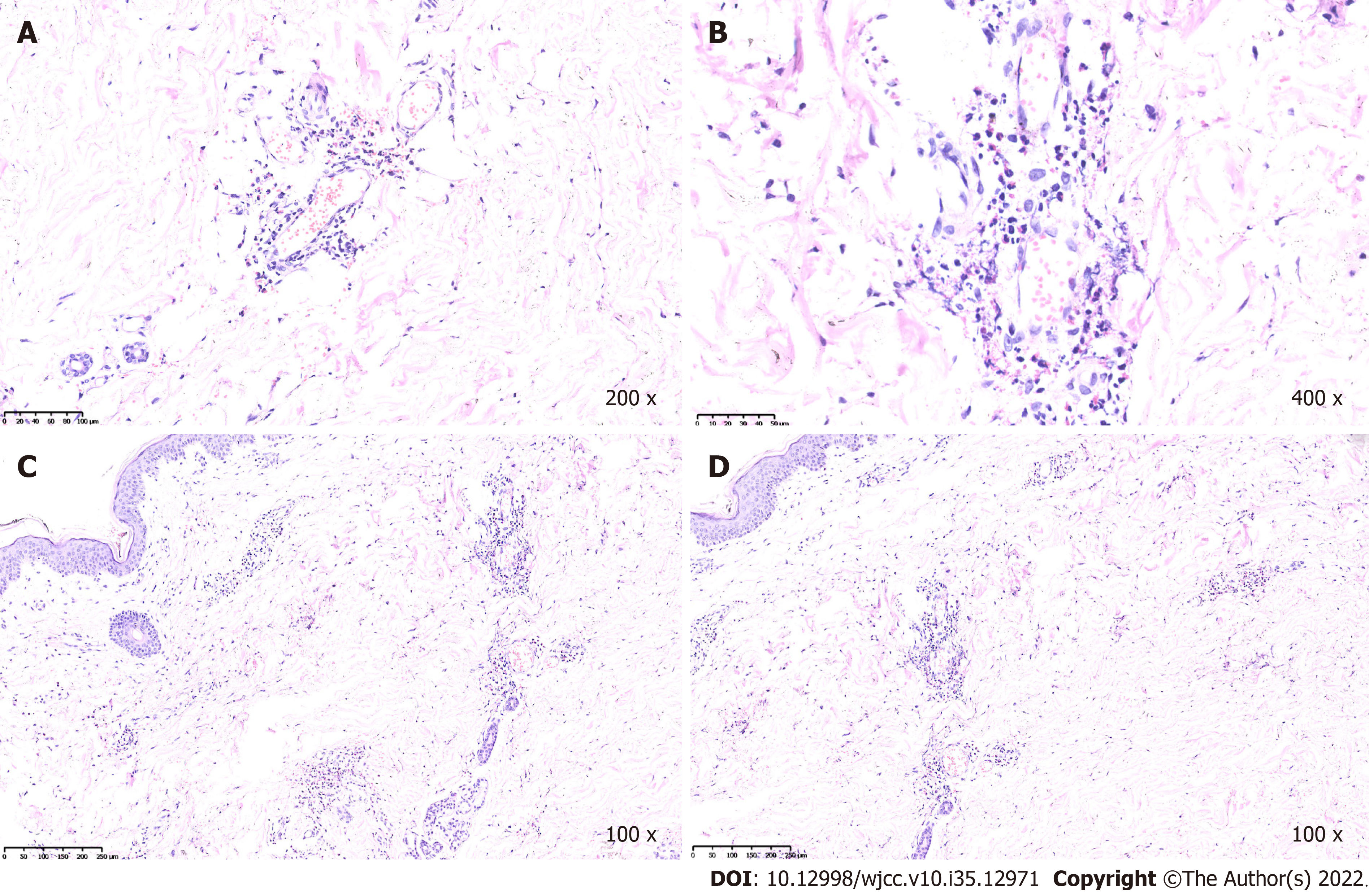
Figure 5 Histological findings of the skin in case 1.
A and B: Representative sections from biopsies of the cutaneous lesions, which revealed the swollen endothelium of small vessels in the dermis, perivascular eosinophils, neutrophils, lymphocytes and broken leukocyte infiltration (hematoxylin and eosin staining); C and D: Representative sections from biopsies, which showed the swollen endothelium of small vessels in the dermis. Perivascular eosinophils, neutrophils, lymphocytes and broken leukocyte infiltration.
- Citation: Li ZG, Zhou JM, Li L, Wang XD. Malignant atrophic papulosis: Two case reports. World J Clin Cases 2022; 10(35): 12971-12979
- URL: https://www.wjgnet.com/2307-8960/full/v10/i35/12971.htm
- DOI: https://dx.doi.org/10.12998/wjcc.v10.i35.12971


