Copyright
©The Author(s) 2022.
World J Clin Cases. Aug 16, 2022; 10(23): 8291-8297
Published online Aug 16, 2022. doi: 10.12998/wjcc.v10.i23.8291
Published online Aug 16, 2022. doi: 10.12998/wjcc.v10.i23.8291
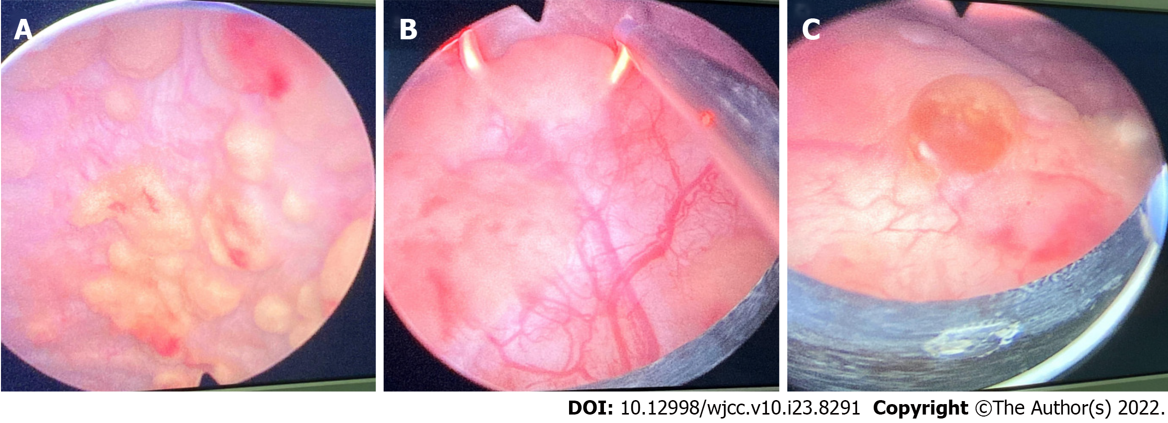
Figure 1 Intraoperative findings.
A: Multiple pale yellow brain loop-like uplifts; B: Tumors being electrocuted; C: Proliferation of scattered inflammatory follicles.
Figure 2 Microscopic findings.
A: Massive Michaelis–Gutman (M-G) bodies can be seen under microscope (Magnification × 200); B: Alkaline M-G bodies arranged in concentric circles as shown by arrows (Magnification × 400); C: Alkaline M-G bodies arranged in concentric circles as shown by arrows (Magnification × 600).
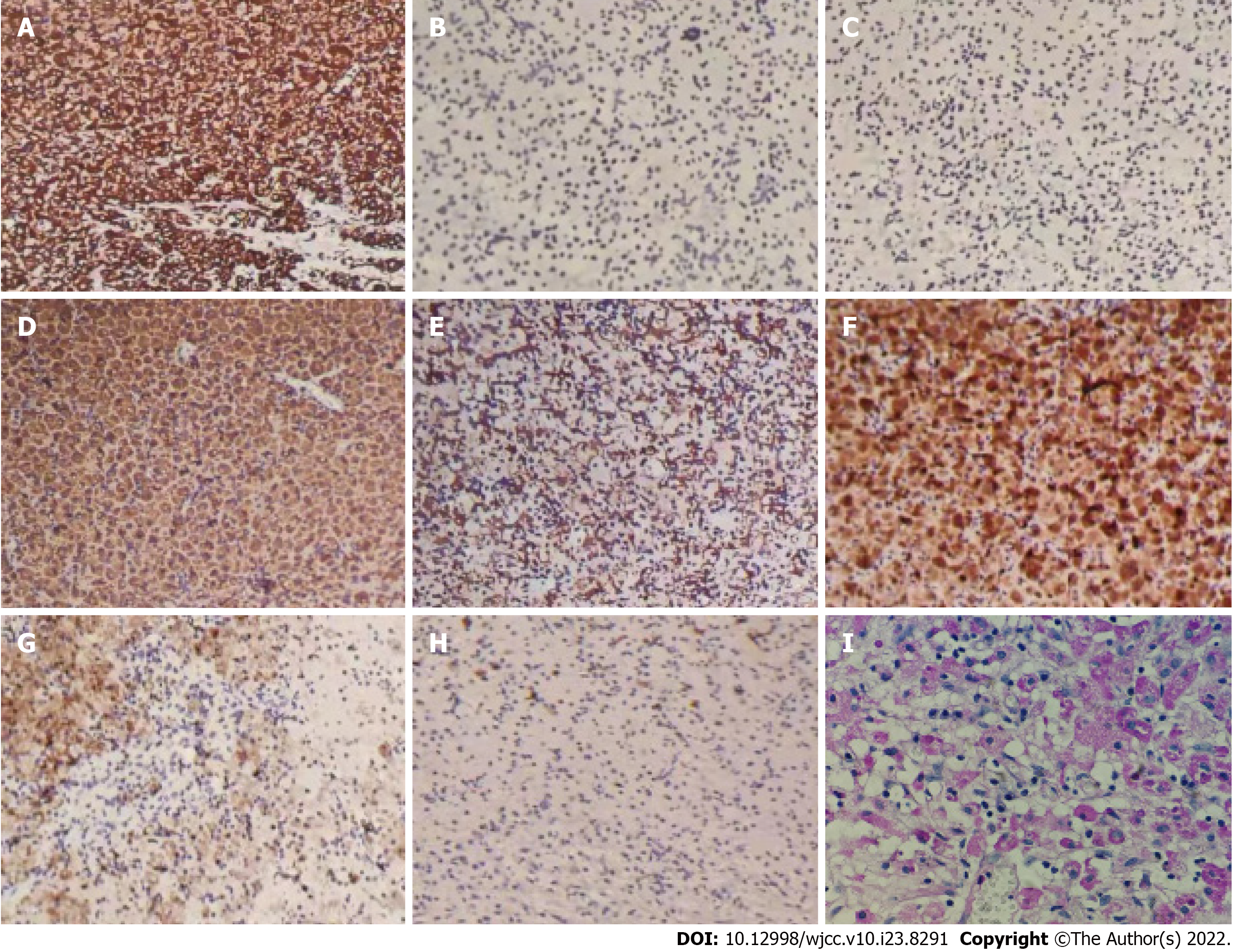
Figure 3 Immunohistochemistry findings.
A: Vimentin (+) (Magnification × 100); B: CKpan (-) (Magnification × 100); C: CK5/6 (-) (Magnification × 100); D: CD68 (+) (Magnification × 100); E: CD163 (+) (Magnification × 100); F: Lysozyme (+) (Magnification × 100); G: MAC387 (-) (Magnification × 100); H: Ki-67 (+2%) (Magnification × 100); I: Michaelis–Gutman body periodic acid-Schiff staining shown in purple red (Magnification × 200).
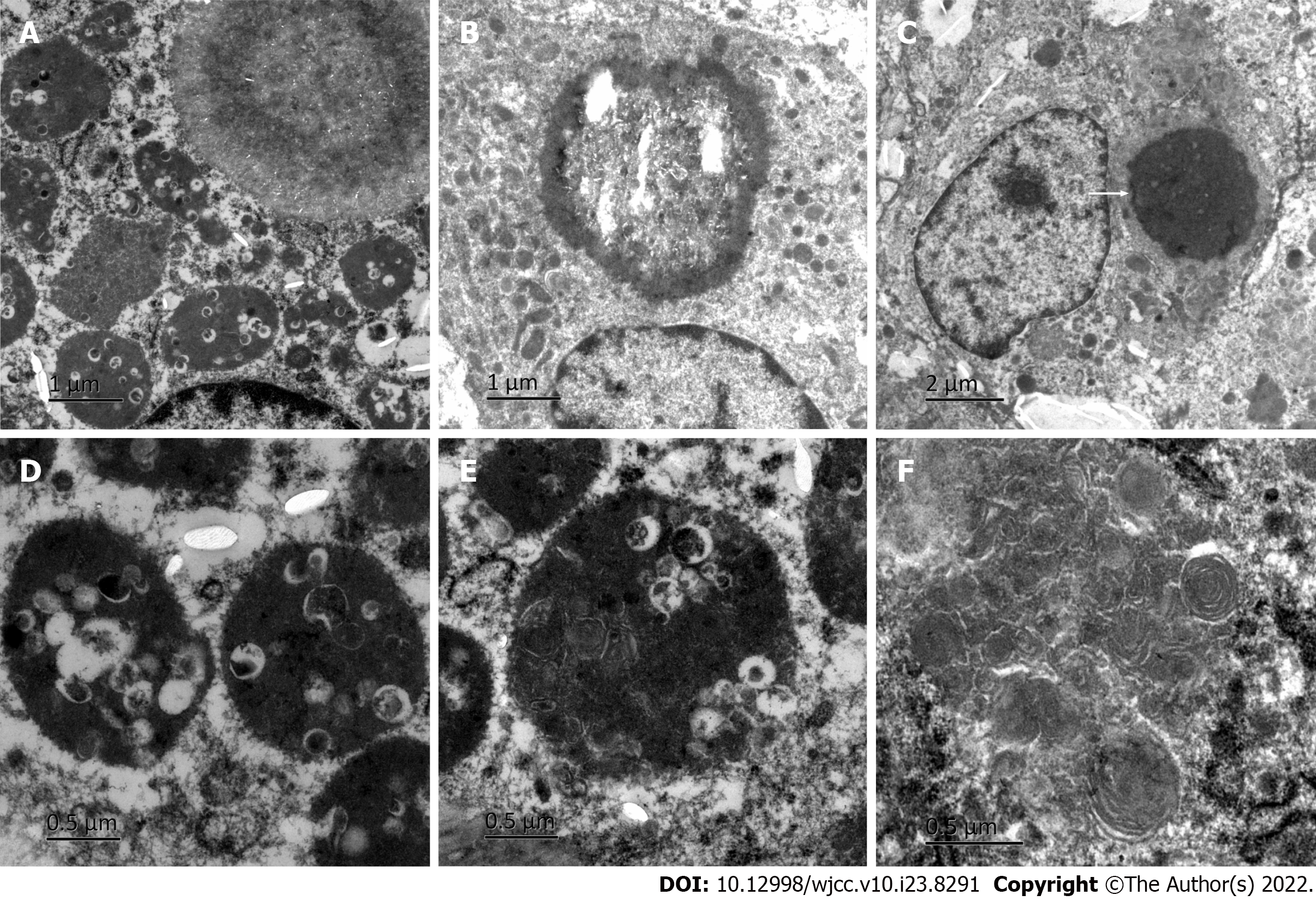
Figure 4 Electron microscopy findings.
A: Phagocytic lysosomes in the cytoplasm of macrophages; B: Microbubble; C: A typical Michaelis–Gutman (M-G) body as shown by arrows; D: Large number of bacteria in phagocytic lysosomes; E: Some bacteria were digested and dissolved; F: Myelin figure.
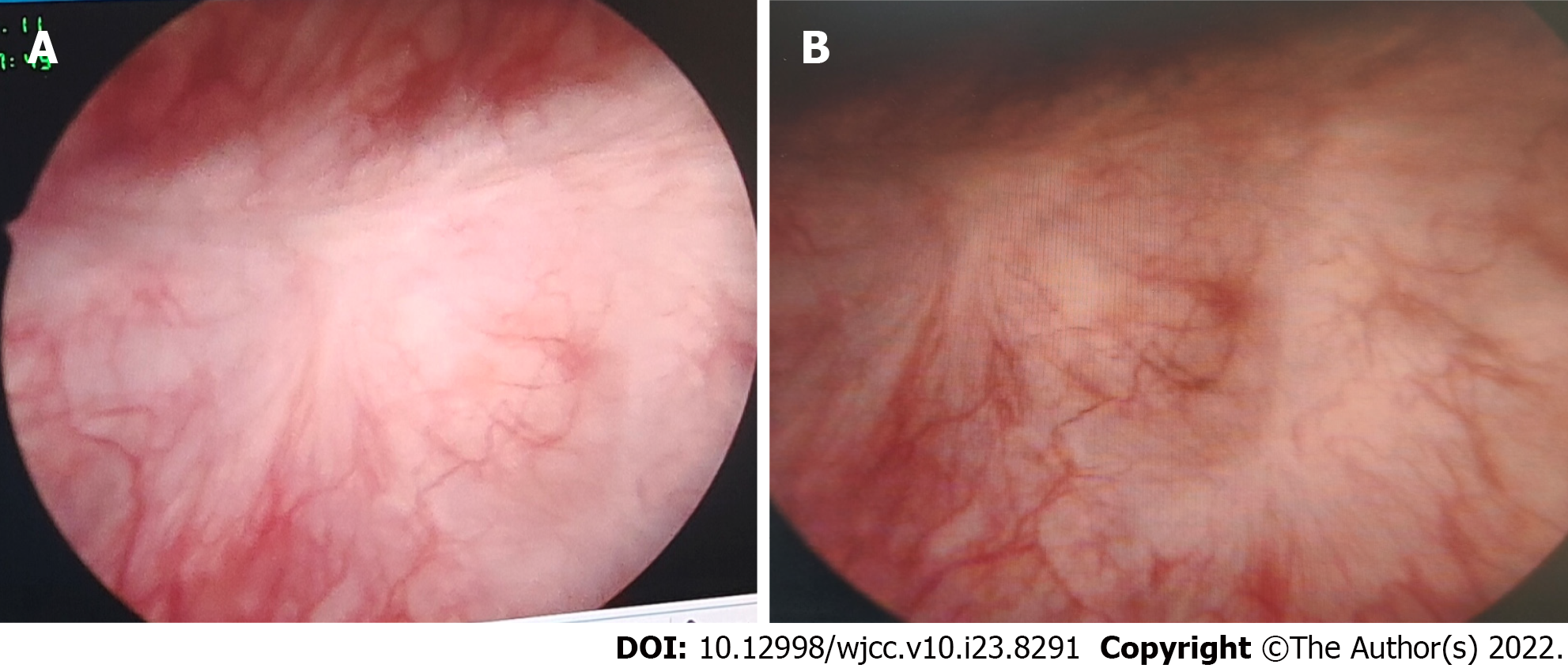
Figure 5 Postoperative review.
A: Scar in the electrotomy area; B: Electrocoagulation scar.
- Citation: Wang HK, Hang G, Wang YY, Wen Q, Chen B. Bladder malacoplakia: A case report. World J Clin Cases 2022; 10(23): 8291-8297
- URL: https://www.wjgnet.com/2307-8960/full/v10/i23/8291.htm
- DOI: https://dx.doi.org/10.12998/wjcc.v10.i23.8291


