Copyright
©The Author(s) 2022.
World J Clin Cases. Jul 6, 2022; 10(19): 6617-6625
Published online Jul 6, 2022. doi: 10.12998/wjcc.v10.i19.6617
Published online Jul 6, 2022. doi: 10.12998/wjcc.v10.i19.6617
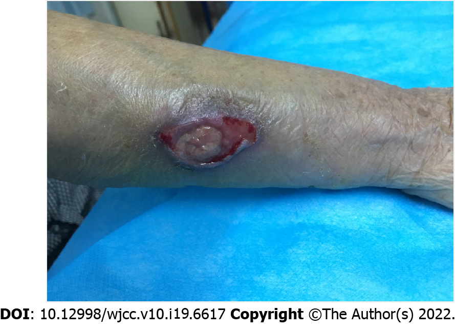
Figure 1 Preoperative clinical photograph of wound ulceration 3 cm × 2 cm in size on the dorsal ulnar aspect of the mid-distal third of right forearm.
The wound partially coated with purulent matter and communicated with the ulnar bone.
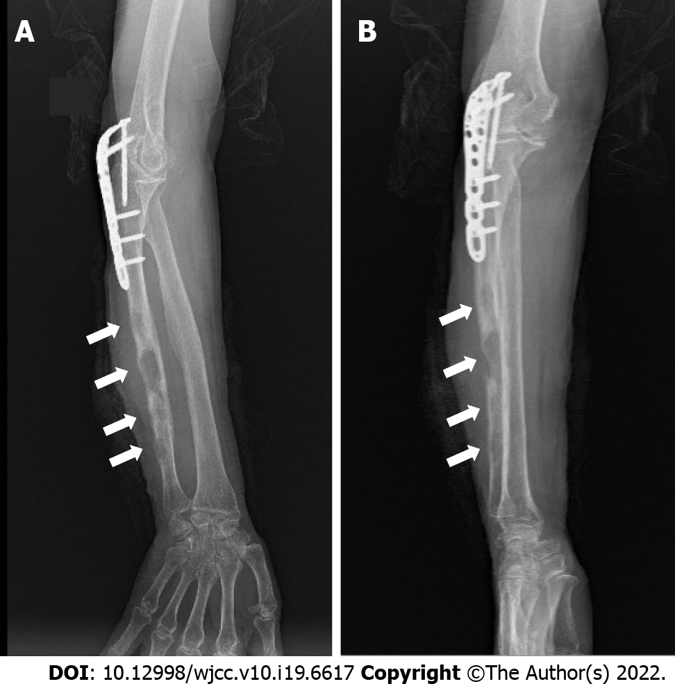
Figure 2 Preoperative X-ray.
A: Anteroposterior film; B: Lateral film. A and B showed multiple irregular low-density osteolytic lesions (white arrows) with little periosteal reaction in the diaphyseal bone at the mid-distal shaft of the ulna, with a swollen soft tissue shadow, the internal fixation of the proximal ulna no loosening.
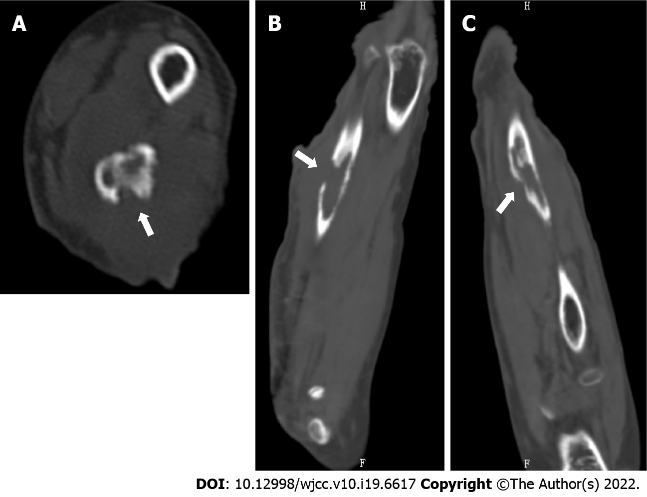
Figure 3 Preoperative computed tomography scan.
A: Horizontal image; B: Coronal image; C: Sagittal image. Images showed the largest bone defect (white arrows) in the mid-distal portion of the right ulna, measuring 2.9 cm × 1.2 cm × 4.2 cm.
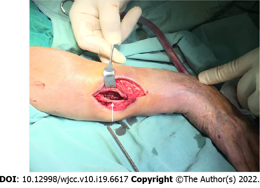
Figure 4 The wound was surgically debrided and irrigated, leaving a bony defect (white arrows) in the right ulna which was filled with amphotericin B-loaded cement.
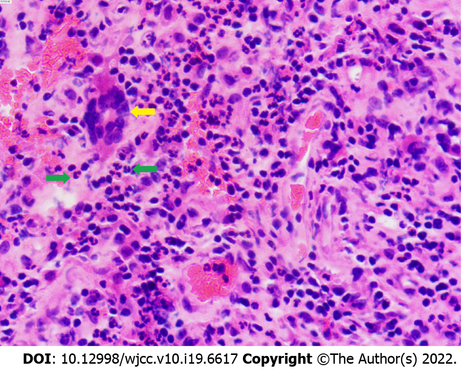
Figure 5 Histopathological examination demonstrated chronic osteomyelitis characterized by granulation tissues with multinucleated giant cells (yellow arrow), and neutrophil infiltration (green arrows) (Hematoxylin-eosin stain 40 × 10).
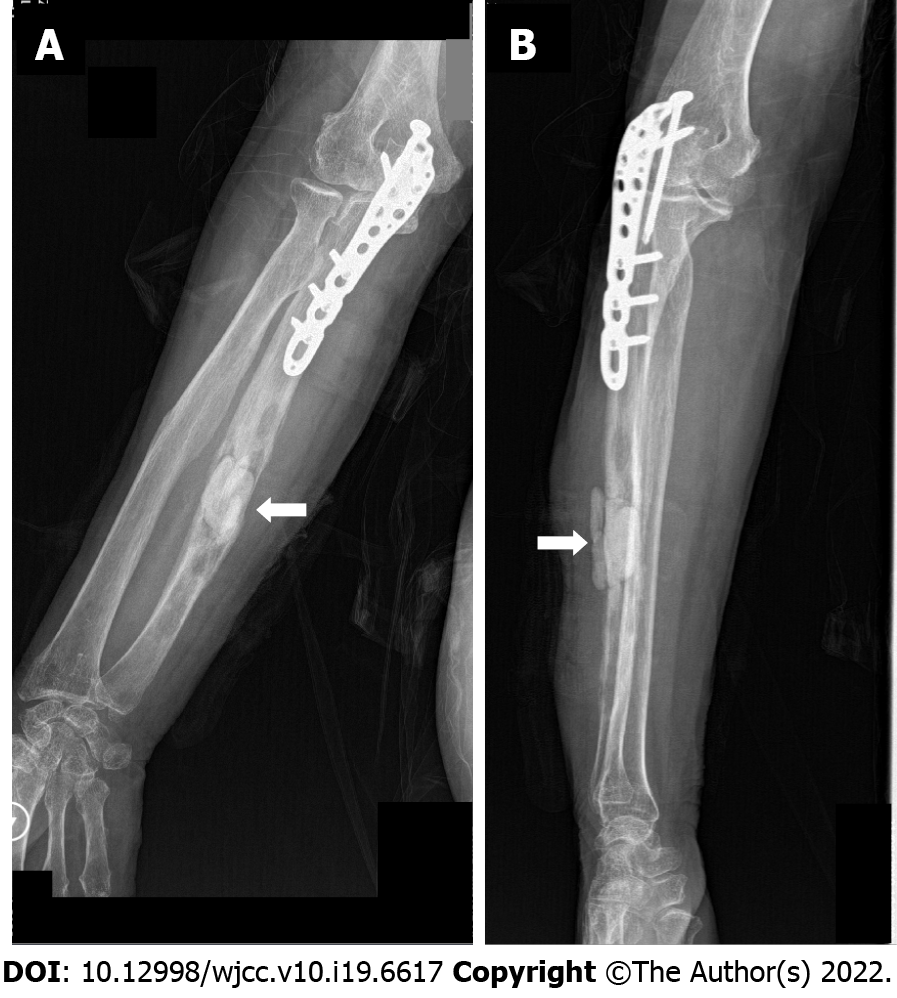
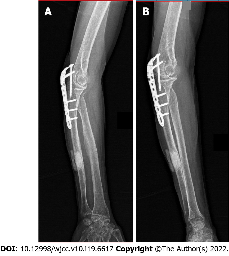
Figure 6 Postoperative X ray.
A: Anteroposterior film; B: Lateral film. A and B showed that the amphotericin B-loaded cement filled the defect area (white arrow) of the right ulna after surgical debridement.
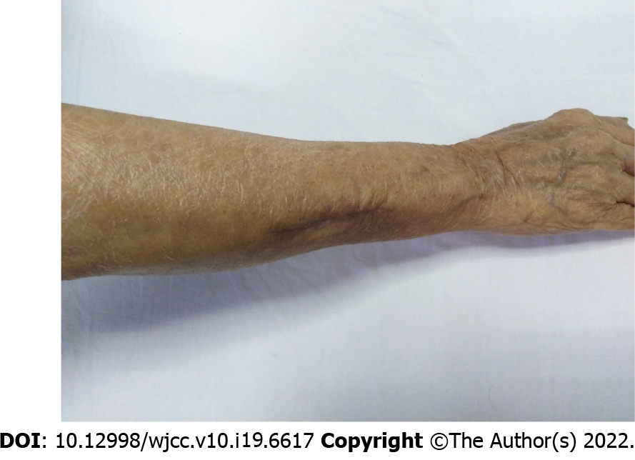
Figure 7 The wound had healed well 3 mo after surgery.
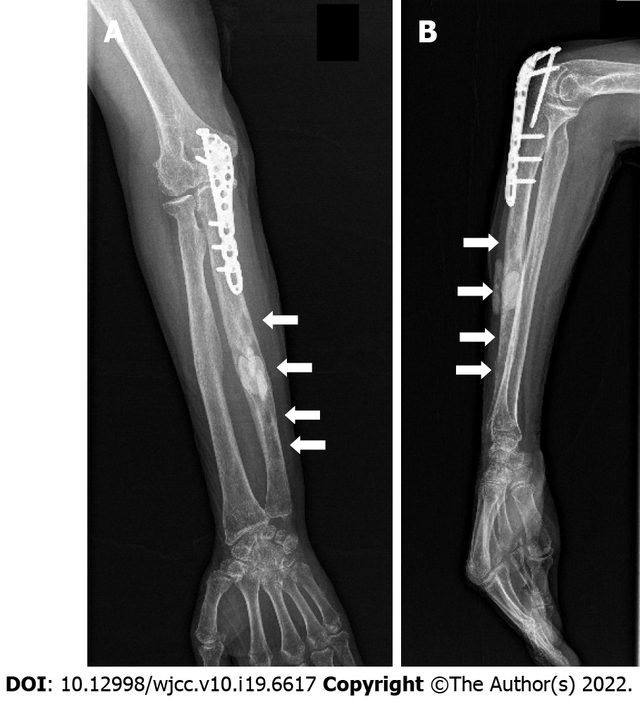
Figure 8 X ray.
A: Anteroposterior film; B: Lateral film. Three months after surgery showed that the osteolytic lesions (white arrows) had shrunk and blurred (except the bone cement filled lesion) in the right ulna.
Figure 9 X ray.
A: Anteroposterior film; B: Lateral film. Ten months after surgery showed the osteolytic lesions had healed (except the bone cement filled lesion) in the right ulna.
- Citation: Ma JL, Liao L, Wan T, Yang FC. Isolated cryptococcal osteomyelitis of the ulna in an immunocompetent patient: A case report. World J Clin Cases 2022; 10(19): 6617-6625
- URL: https://www.wjgnet.com/2307-8960/full/v10/i19/6617.htm
- DOI: https://dx.doi.org/10.12998/wjcc.v10.i19.6617


