
















©The Author(s) 2022.
World J Clin Cases. Jul 6, 2022; 10(19): 6617-6625
Published online Jul 6, 2022. doi: 10.12998/wjcc.v10.i19.6617
Published online Jul 6, 2022. doi: 10.12998/wjcc.v10.i19.6617
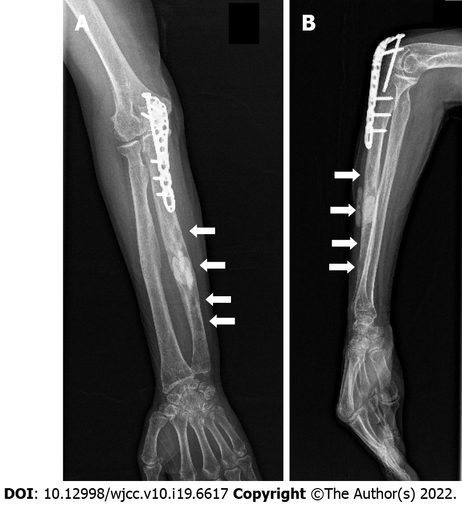
Figure 8 X ray.
A: Anteroposterior film; B: Lateral film. Three months after surgery showed that the osteolytic lesions (white arrows) had shrunk and blurred (except the bone cement filled lesion) in the right ulna.
- Citation: Ma JL, Liao L, Wan T, Yang FC. Isolated cryptococcal osteomyelitis of the ulna in an immunocompetent patient: A case report. World J Clin Cases 2022; 10(19): 6617-6625
- URL: https://www.wjgnet.com/2307-8960/full/v10/i19/6617.htm
- DOI: https://dx.doi.org/10.12998/wjcc.v10.i19.6617