Copyright
©The Author(s) 2025.
World J Psychiatry. Aug 19, 2025; 15(8): 107342
Published online Aug 19, 2025. doi: 10.5498/wjp.v15.i8.107342
Published online Aug 19, 2025. doi: 10.5498/wjp.v15.i8.107342
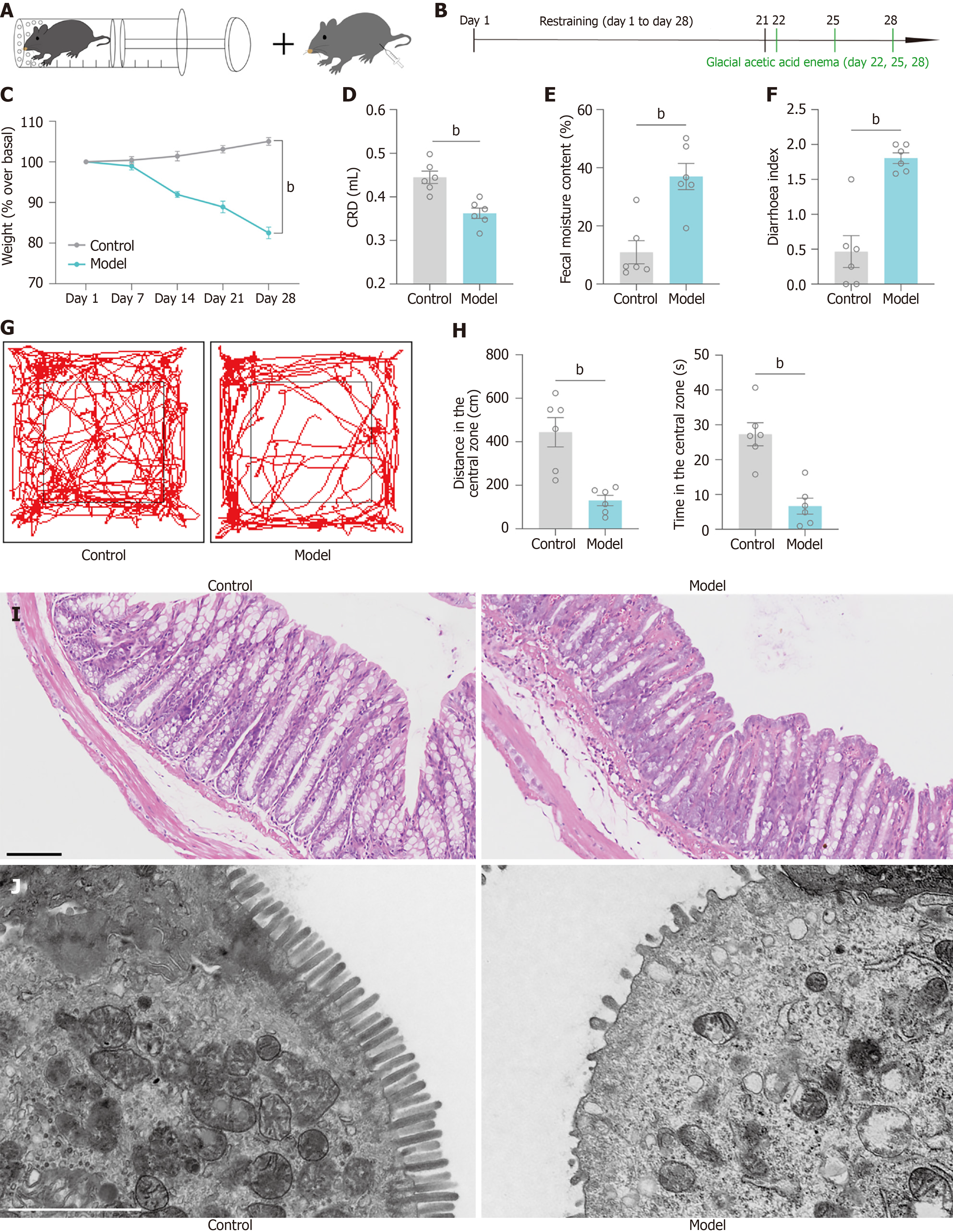
Figure 1 Chronic restraint combined with glacial acetic acid enema was used to induce diarrhoeal irritable bowel syndrome accompanied by negative emotions.
A: Schematic diagram of chronic restraint combined with glacial acetic acid enema; B: Flow diagram of chronic restraint combined with glacial acetic acid enema; C: Statistical graphs of the changes in the body weights of mice at different time points; D: Statistical graph of the amount of saline injected for the assessment of the colorectal dilatation-induced abdominal withdrawal reflex; E: Statistical graph of the faecal water content in mice; F: Statistical graph of the diarrhoea indices of the mice; G: Paths travelled by mice in the open-field test; H: Statistical plots of distance in center and time in center by mice in the open-field test; I: Pathological changes in the colonic mucosa of model mice were observed by Hematoxylin eosin staining. Scale bar, 100 μm; J: Alterations in the colonic villi of model mice observed by transmission electron microscopy. Scale bar, 2.0 μm; n = 6 mice in each group, model group vs control group; aP < 0.05, bP < 0.01.
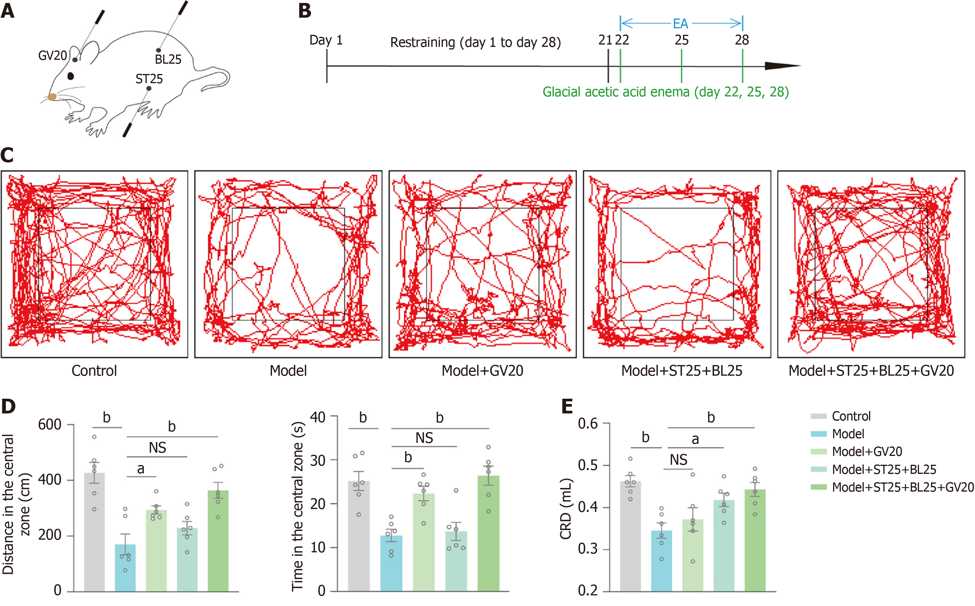
Figure 2 Effects of electroacupuncture at different acupoints on diarrhoeal irritable bowel syndrome.
A: Schematic representation of the electroacupuncture positions of Baihui (GV20), Tianshu (ST25) and Dachangshu (BL25); B: Flowchart diagram of electroacupuncture acupoints in mice; C: Paths travelled by the mice in each group in the open-field test; D: Statistical graphs of distance in center and time in center of the mice in the open-field test; E: Statistical graph of saline injections in each group of mice in the colorectal dilatation test; n = 6 mice in each group, model group vs control group, model + GV20/model + ST25 + BL25/model + ST25 + BL25 + GV20 group vs model group; aP < 0.05, bP < 0.01; NS: Not significant; GV20: Baihui; ST25: Tianshu; BL25: Dachangshu; EA: Electroacupuncture.
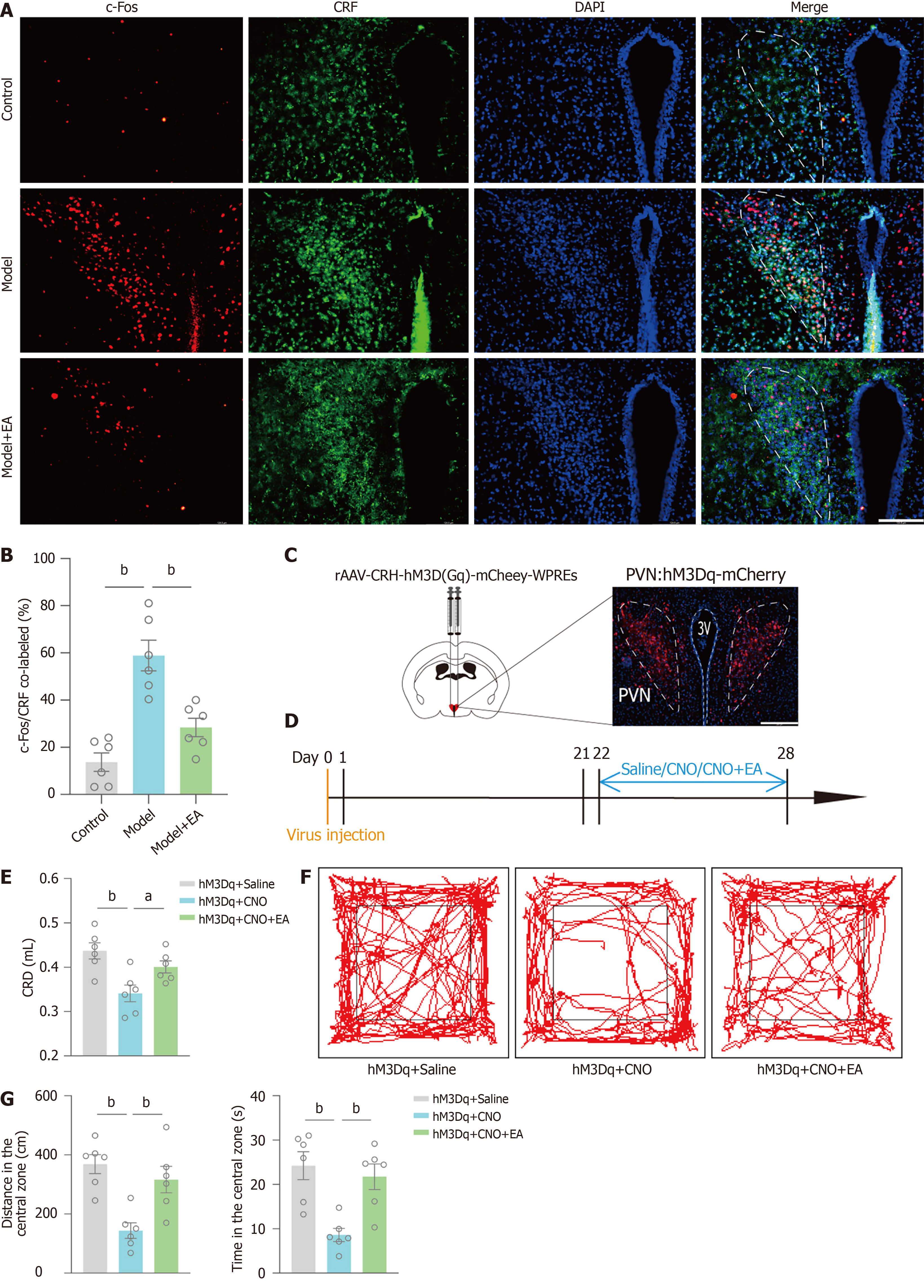
Figure 3 Corticotropin-releasing factor-ergic neurons of the paraventricular hypothalamic nucleus participate in the effect of electroacupuncture.
A: Immunofluorescence plots of the paraventricular hypothalamic nucleus (PVN) in the control group, model group and model + electroacupuncture (EA) (Baihui + Tianshu + Dachangshu) group [c-fos is red, Corticotropin-releasing factor (CRF) is green, and 4,6-diamidino-2-phenylindole is blue]. Scale bar, 124.5 μm; B: Statistical plot of the c-fos and CRF co-labelling rates in the PVN. Each group n = 6, model group vs control group, model + EA group vs model group, bP < 0.01; C: The virus (red fluorescence) was expressed in the PVN. Scale bar, 249 μm; D: Injection of the chemogenetically activated virus rAAV-CRH-hM3D(Gq)-mCherry-WPREs into bilateral PVNs and intraperitoneal injection of saline, clozapine-N-oxide (CNO), or CNO plus EA after 21 days; E: Statistical graphs of the amount of saline injected into the mice in the colorectal dilatation assay; F: Paths travelled by mice in each group in the open-field test; G: Statistical graphs of distance in center and time in center by mice in the open-field test; n = 6 mice in each group, hM3Dq + CNO group vs hM3Dq + saline group, hM3Dq + CNO + EA group vs hM3Dq + CNO group; aP < 0.05, bP < 0.01; CRF: Corticotropin-releasing factor; PVN: Paraventricular hypothalamic nucleus; EA: Electroacupuncture; CNO: Clozapine-N-oxide; DAPI: 4,6-diamidino-2-phenylindole.
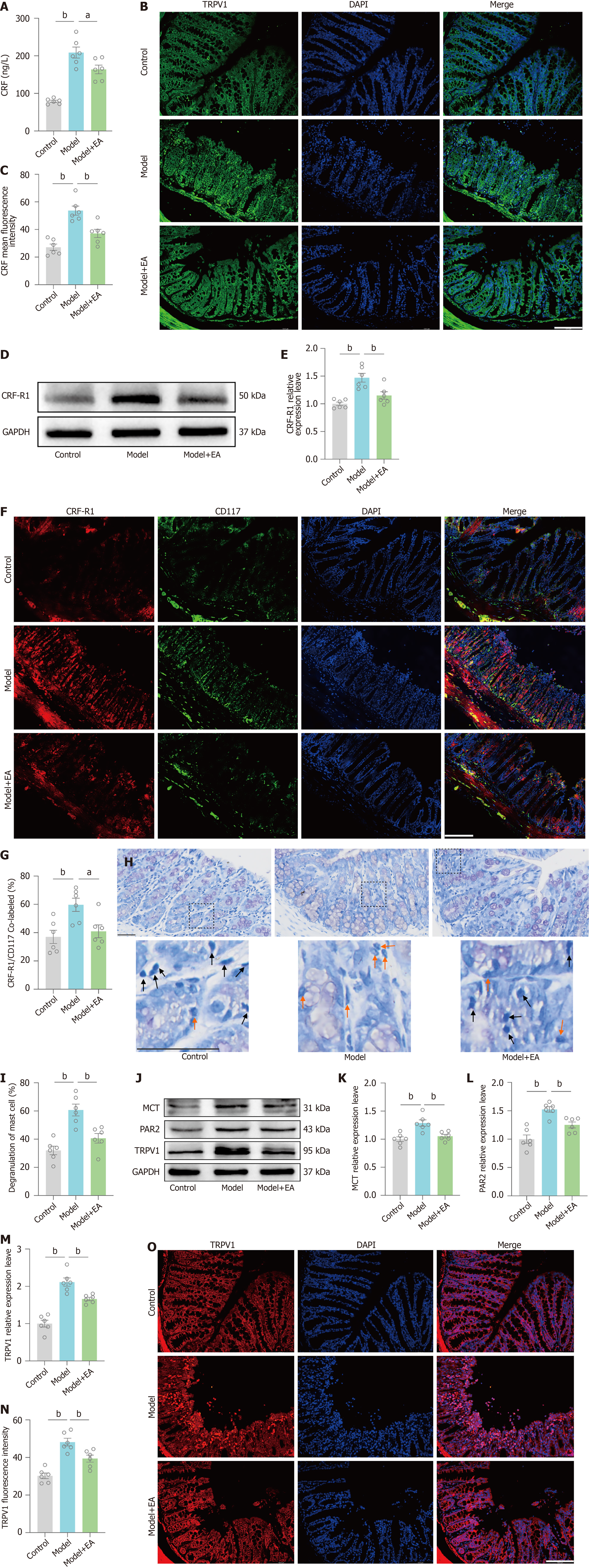
Figure 4 The peripheral corticotropin-releasing factor-colonic mast cell/transient potential receptor vanilloid type 1 pathway is involved in the therapeutic effects of electroacupuncture.
A: Statistical graph of the serum corticotropin-releasing factor (CRF) levels detected by enzyme-linked immunosorbent assay in each group of mice; B: Representative plots of CRF in the colon of each group of mice determined by an immunofluorescence assay. Scale bar, 124.5 μm; C: Statistical graph of the average fluorescence intensity of CRF expression levels in the colons of the mice in each group using immunofluorescence detection; D: Representative image of the Western blot of corticotropin-releasing factor-receptor 1 (CRF-R1); E: Statistical graph of the relative expression level of colonic CRF-R1 detected by Western blot in each group of mice; F: Immunofluorescence plots in the colons of each group of mice. (CRF-R1 is red, CD117 is green, and 4,6-diamidino-2-phenylindole is blue). Scale bar, 124.5 μm; G: Statistical plot of the CRF-R1 and CD117 co-labelling rates in the colons of the mice in each group; H: Mast cell (MC) degranulation rate of the colon in each group of mice detected by toluidine blue staining. Black arrows represent steady state MCs, orange arrows represent degranulated MCs. Scale bar, 50 μm; I: Statistical graph of the MC degranulation rate in each group of mice; J: Representative image of the Western blot of MC tryptase (MCT), protease-activated receptor 2 (PAR2) and transient potential receptor vanilloid type 1 (TRPV1) in mice colons were detected by Western blot; K: Statistical graph of the relative expression levels of MCT; L: Statistical graph of the relative expression levels of PAR2; M: Statistical graph of the relative expression levels of TRPV1; N: Statistical graph of the average fluorescence intensity of TRPV1 expression levels in the colons of the mice in each group; O: Representative plots of TRPV1 in the colon of each group of mice following an immunofluorescence assay. Scale bar, 124.5 μm; Each group n = 6, model group vs control group, model + EA group vs model group; aP < 0.05, bP < 0.01; CRF: Corticotropin-releasing factor; MC: Mast cell; TRPV1: Transient potential receptor vanilloid type 1; CRF-R1: Corticotropin-releasing factor-receptor 1; MCT: Mast cell tryptase; PAR2: Protease-activated receptor 2; EA: Electroacupuncture; DAPI: 4,6-diamidino-2-phenylindole.
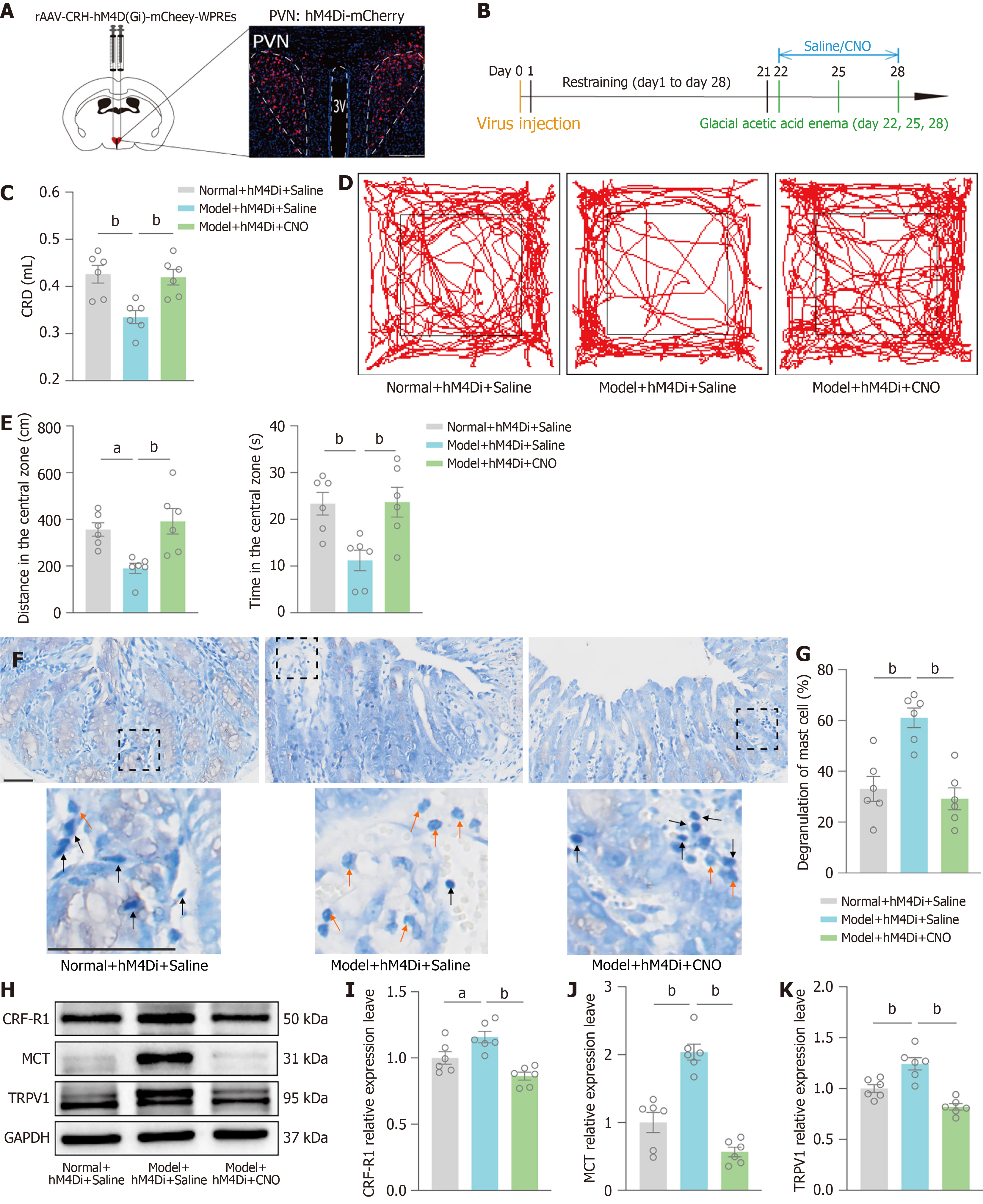
Figure 5 Corticotropin-releasing factor-ergic neurons of the paraventricular hypothalamic nucleus may regulate negative emotions and visceral hypersensitivity through colonic corticotropin-releasing factor-mast cell/transient potential receptor vanilloid type 1 pathway.
A: Viruses (red fluorescence) were expressed on paraventricular hypothalamic nucleus (PVNs). Scale bar, 249 μm; B: Injection of the chemogenetically inhibited virus rAAV-CRH-hM4D(Gi)-mCherry-WPREs into bilateral PVNs and intraperitoneal injection of saline/clozapine-N-oxide (CNO) 21 days later; C: Statistical graph of saline injections in mice in the colorectal dilatation test; D: Paths travelled by mice in the open-field test; E: Statistical graphs of distance in center and time in center by mice in the open-field test; F: Detection of the mast cell (MC) degranulation rate in the mouse colon by toluidine blue staining in each group. Black arrows represent steady state MCs, orange arrows represent degranulated MCs. Scale bar, 50 μm; G: Statistical graphs of the MC degranulation rate; H: Representative image of the Western blot of corticotropin-releasing factor-receptor 1 (CRF-R1), mast cell tryptase (MCT) and transient potential receptor vanilloid type 1 (TRPV1) in mice colons were detected by Western blot; I: Statistical graph of the relative expression levels of CRF-R1; J: Statistical graph of the relative expression levels of MCT; K: Statistical graph of the relative expression levels of TRPV1; Each group n = 6, model + hM4Di + saline group vs normal + hM4Di + saline group, model + hM4Di + CNO group vs model + hM4Di + saline group; aP < 0.05, bP < 0.01; CRF: Corticotropin-releasing factor; PVN: Paraventricular hypothalamic nucleus; MC: Mast cell; TRPV1: Transient potential receptor vanilloid type 1; CNO: Clozapine-N-oxide; CRF-R1: Corticotropin-releasing factor-receptor 1; MCT: Mast cell tryptase.
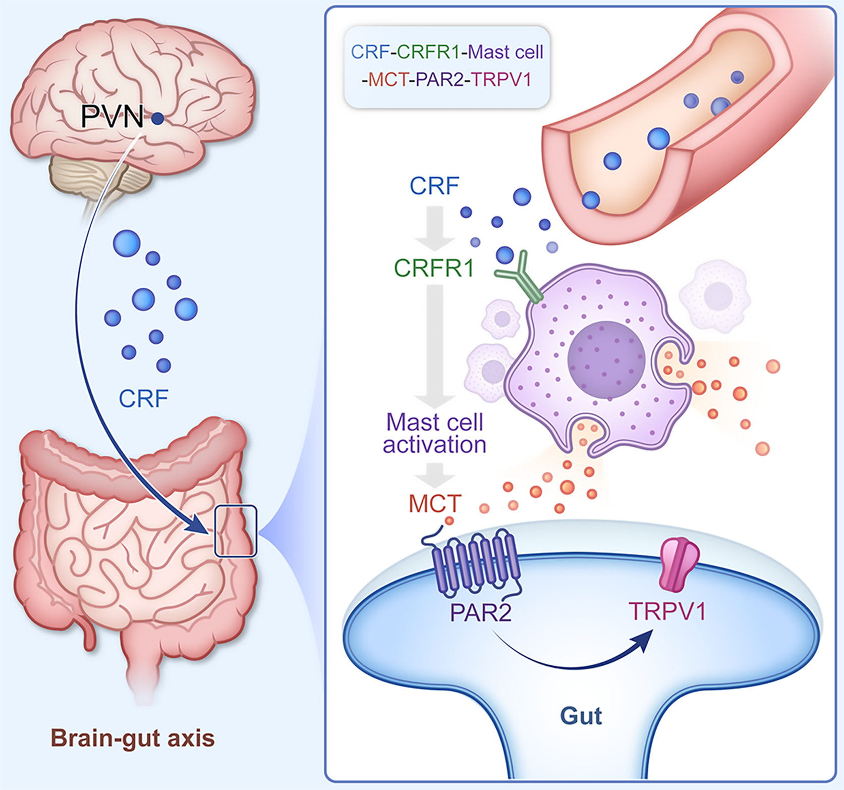
Figure 6 Graphical abstract.
Brain-colon crosstalk may mediate the pathological process of diarrhoeal irritable bowel syndrome and the mechanism of action of electroacupuncture. We propose that chronic restraint combined with glacial acetic acid enema activated paraventricular hypothalamic nucleus corticotropin-releasing factor (CRF)-ergic neurons, increased peripheral CRF, increased CRF-receptor 1 in colonic mast cells, resulted in mast cell degranulation, and then upregulated protease-activated receptor 2 and transient potential receptor vanilloid 1 in gut neurons, which ultimately induced negative emotions and visceral hypersensitivity; however, electroacupuncture reversed this phenomenon. CRF: Corticotropin-releasing factor.
- Citation: Xu JG, Yuan Y, Ma HK, Huang S, Zhu SL, Li TT, Wang XY, Shen GM, Wang H. Electroacupuncture ameliorates visceral hypersensitivity and negative emotions by regulating paraventricular hypothalamic nucleus and colonic corticotropin-releasing factor signaling. World J Psychiatry 2025; 15(8): 107342
- URL: https://www.wjgnet.com/2220-3206/full/v15/i8/107342.htm
- DOI: https://dx.doi.org/10.5498/wjp.v15.i8.107342


