Copyright
©2012 Baishideng Publishing Group Co.
World J Radiol. Aug 28, 2012; 4(8): 345-352
Published online Aug 28, 2012. doi: 10.4329/wjr.v4.i8.345
Published online Aug 28, 2012. doi: 10.4329/wjr.v4.i8.345
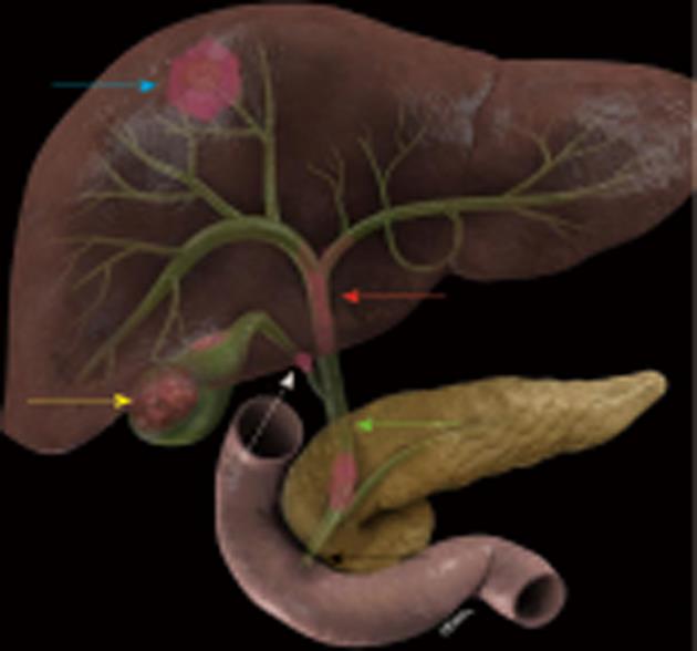
Figure 1 Anatomical distribution of cholangiocarcinoma.
The intrahepatic cholangiocarcinoma present most as solid masses in the liver (blue arrow), gallbladder (GB) cancer present as solid mass in the GB (yellow arrow), proximal bile duct tumors present mostly as infiltrating masses (red arrow), middle bile duct tumors (green arrow), and distal bile duct tumors (black arrow). The cystic duct is labeled with a white arrow.
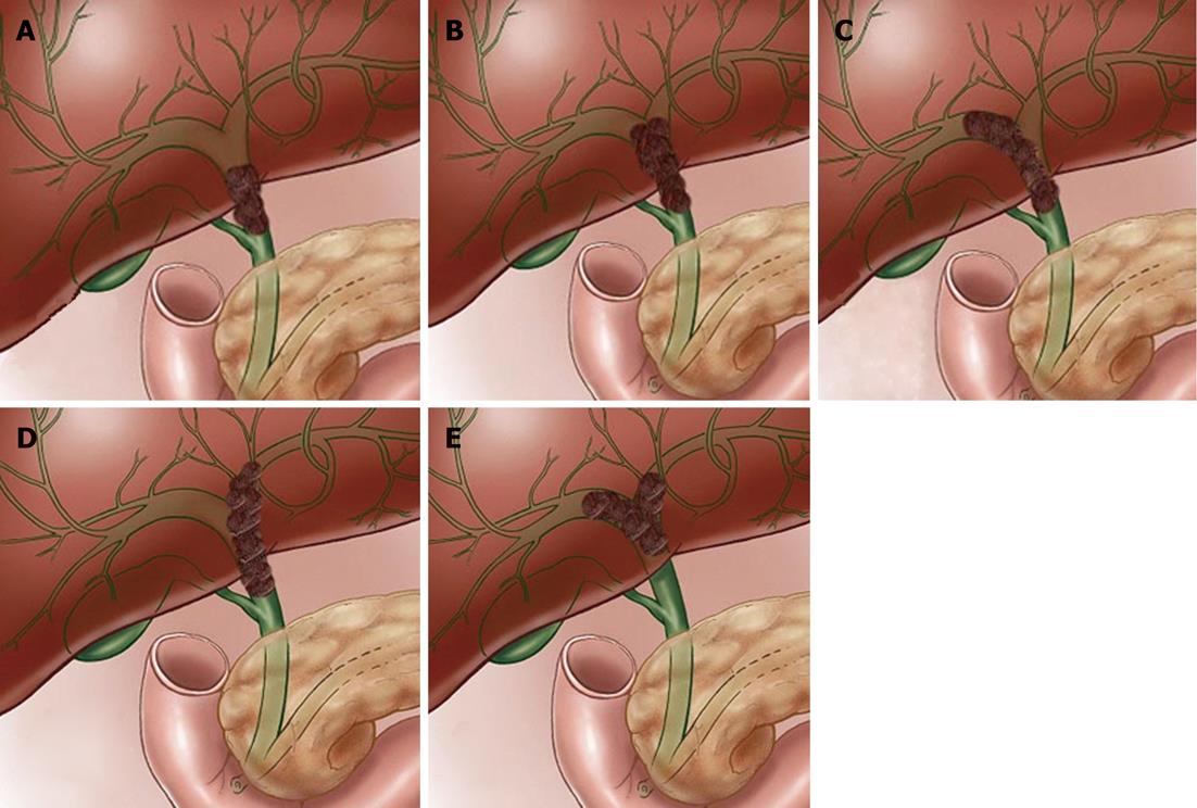
Figure 2 Hilar tumor classification.
A: Type I hilar tumors are proximal bile duct tumors that do not extend to the bifurcation; B: Type II tumors extend to the bifurcation without extension into the intrahepatic bile ducts; C:Type IIIa proximal bile duct tumors correspond to tumors that extend to the right intrahepatic bile ducts; D: Type IIIb proximal bile duct tumors correspond to tumors that extend to the left intrahepatic bile ducts; E: Type IV tumors extend to the right and left intrahepatic bile ducts.
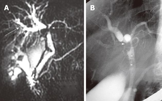
Figure 3 Bile duct tumor.
A: Magnetic resonance cholangiopancreatography (MRCP) of a 57-year-old female with a proximal bile duct tumor (dash arrow). There is dilatation of the right intrahepatic bile ducts (solid arrow); B: Corresponding endoscopic cholangiopancreatography of 57-year-old female with proximal bile duct tumor (arrowhead). This is a type IIIa tumor as per Bismouth-Corlette classification.
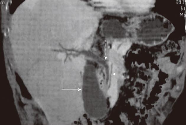
Figure 4 Computed tomography cholangiopancreatography utilizing minimum intensity projection.
The common bile duct (dash arrow), the pancreatic duct (arrowhead), and the gallbladder (solid arrow) are visualized and are normal in appearance.
Figure 5 T-staging for proximal bile duct tumors.
A: T1 tumors are confined to the bile duct wall without extension to the periductal fat; B: The T2 tumors extend to the periductal fat or the liver; C: T3 tumors have unilateral extension to the portal vein (arrow) or hepatic artery; D: T4 tumors extend to the main portal vein (arrow), common hepatic artery, secondary biliary radicles, or contralateral vascular extension.
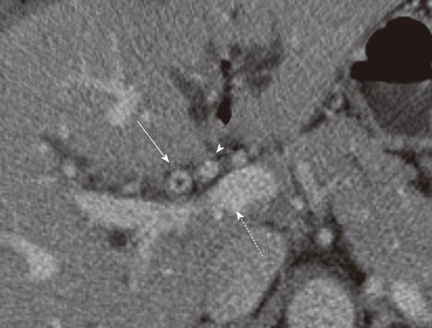
Figure 6 Proximal bile tumor T2 stage.
Post-contrast computed tomography exam at the level of the common hepatic duct in a 62-year-old male with biliary cancer. There is enhancement and thickening of the bile duct wall (solid arrow). The fat around the duct is not preserved but the portal vein (dash arrow) and hepatic artery (arrowhead) are spared. Radiologically, this is a T2 tumor due to involvement of periductal fat.
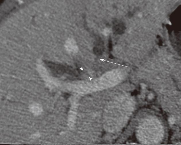
Figure 7 T4 - proximal bile tumor.
Post-contrast computed tomography exam at the level of the intrahepatic bile duct in a 58-year-old female with biliary cancer. There is enhancement and thickening of the right intrahepatic bile duct wall (arrowheads). There is abrupt termination of the left intrahepatic bile ducts (arrow) in keeping with tumor extension). Radiologically, this is a T4 tumor due to bilateral involvement of secondary biliary radicles
Figure 8 T-Staging for distal bile duct tumors.
A: T1 tumors are confined to the bile duct wall without extension to the periductal fat; B: T2 tumors extend to the periductal fat or the liver; C: T3 tumors have adjacent organs without celiac axis or superior mesenteric artery invasion; D:T4 tumors extend to the superior mesenteric artery (arrow) or celiac artery.
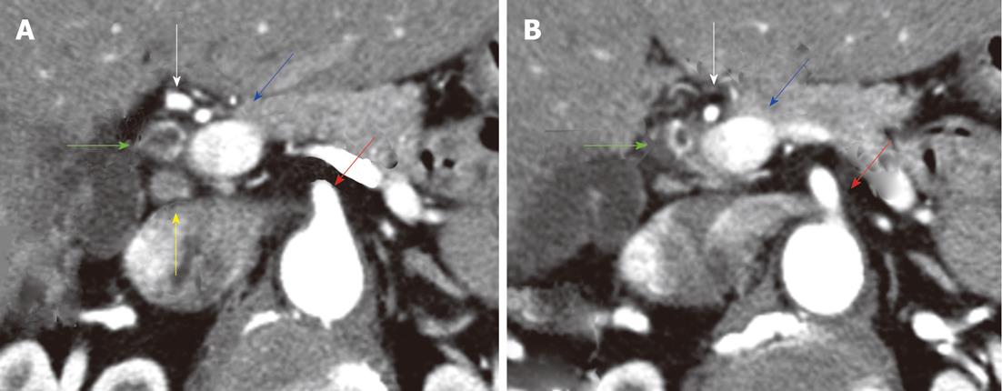
Figure 9 Distal bile duct tumors.
Post-contrast computed tomography (CT) examination of the abdomen in a 58-year-old female with cholangiocarcinoma of the bile duct; A: CT image at a cranial location shows dilatation of the bile duct and bile duct enhancement (green arrow); B: CT image at a caudal location shows enhancement of the distal bile duct. There is a small regional lymph node (yellow arrow). The hepatic artery (white arrow), portal vein (blue arrow), and superior mesenteric artery (red arrow) are spared. Radiologically, appearances are consistent with a T1 tumor (periductal fat not involved) and this was confirmed by pathology.
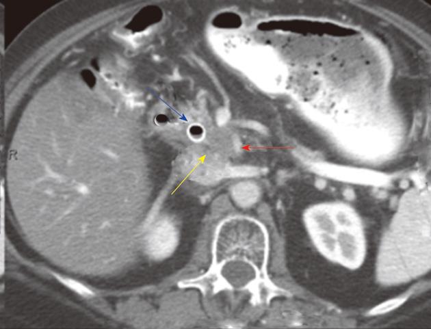
Figure 10 Distal bile duct tumors.
Post-contrast computed tomography (CT) examination of the abdomen in a 65-year-old female with cholangiocarcinoma of the bile duct. CT image of the distal bile at the level of the pancreas shows a soft tissue tumor (yellow arrow) in keeping with the tumor. The tumor extends to the superior mesenteric artery (red arrow). There is a biliary stent in place (blue arrow). Radiologically, appearances are consistent with a T4 tumor due to involvement of superior mesenteric artery.
- Citation: Ganeshan D, Moron FE, Szklaruk J. Extrahepatic biliary cancer: New staging classification. World J Radiol 2012; 4(8): 345-352
- URL: https://www.wjgnet.com/1949-8470/full/v4/i8/345.htm
- DOI: https://dx.doi.org/10.4329/wjr.v4.i8.345


