Copyright
©2012 Baishideng Publishing Group Co.
World J Radiol. Aug 28, 2012; 4(8): 345-352
Published online Aug 28, 2012. doi: 10.4329/wjr.v4.i8.345
Published online Aug 28, 2012. doi: 10.4329/wjr.v4.i8.345
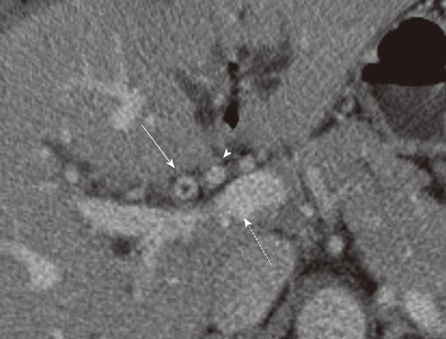
Figure 6 Proximal bile tumor T2 stage.
Post-contrast computed tomography exam at the level of the common hepatic duct in a 62-year-old male with biliary cancer. There is enhancement and thickening of the bile duct wall (solid arrow). The fat around the duct is not preserved but the portal vein (dash arrow) and hepatic artery (arrowhead) are spared. Radiologically, this is a T2 tumor due to involvement of periductal fat.
- Citation: Ganeshan D, Moron FE, Szklaruk J. Extrahepatic biliary cancer: New staging classification. World J Radiol 2012; 4(8): 345-352
- URL: https://www.wjgnet.com/1949-8470/full/v4/i8/345.htm
- DOI: https://dx.doi.org/10.4329/wjr.v4.i8.345


