Copyright
©2012 Baishideng Publishing Group Co.
World J Radiol. Aug 28, 2012; 4(8): 345-352
Published online Aug 28, 2012. doi: 10.4329/wjr.v4.i8.345
Published online Aug 28, 2012. doi: 10.4329/wjr.v4.i8.345
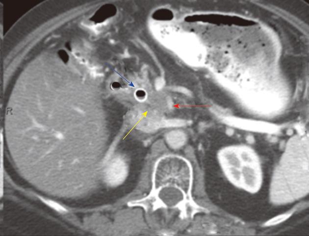
Figure 10 Distal bile duct tumors.
Post-contrast computed tomography (CT) examination of the abdomen in a 65-year-old female with cholangiocarcinoma of the bile duct. CT image of the distal bile at the level of the pancreas shows a soft tissue tumor (yellow arrow) in keeping with the tumor. The tumor extends to the superior mesenteric artery (red arrow). There is a biliary stent in place (blue arrow). Radiologically, appearances are consistent with a T4 tumor due to involvement of superior mesenteric artery.
- Citation: Ganeshan D, Moron FE, Szklaruk J. Extrahepatic biliary cancer: New staging classification. World J Radiol 2012; 4(8): 345-352
- URL: https://www.wjgnet.com/1949-8470/full/v4/i8/345.htm
- DOI: https://dx.doi.org/10.4329/wjr.v4.i8.345


