Copyright
©The Author(s) 2025.
World J Radiol. Aug 28, 2025; 17(8): 111065
Published online Aug 28, 2025. doi: 10.4329/wjr.v17.i8.111065
Published online Aug 28, 2025. doi: 10.4329/wjr.v17.i8.111065
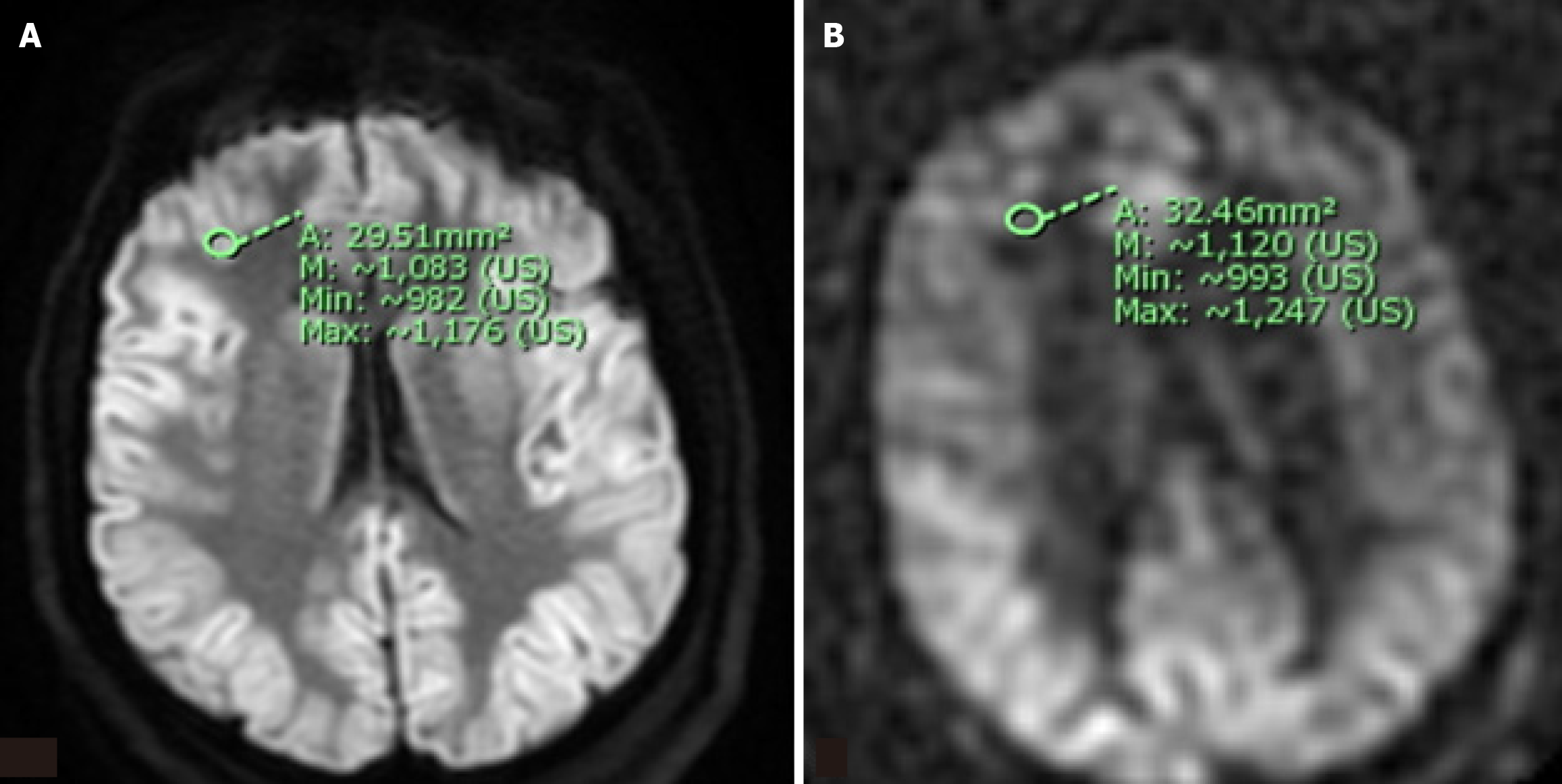
Figure 1 Representative diffusion weighted imaging and arterial spin-labeling sequences illustrating region of interest selection for semiquantiative analysis of signal intensity.
A region of interest (ROI) measuring approximately 30 mm² areas was selected for each of twelve brain regions (bilateral frontal, parietal, temporal, and occipital lobes; thalami; and basal ganglia). The raw mean and maximum signal intensity within each ROI was recorded and normalized on a 0-12 point scale using the signal intensity within the ipsilateral cerebellar hemisphere as an internal reference value. A: A representative example of an ROI within the right frontal lobe on a diffusion weighted imaging sequence; B: A representative example of an ROI within the right frontal lobe on the corresponding arterial spin-labeling sequence.
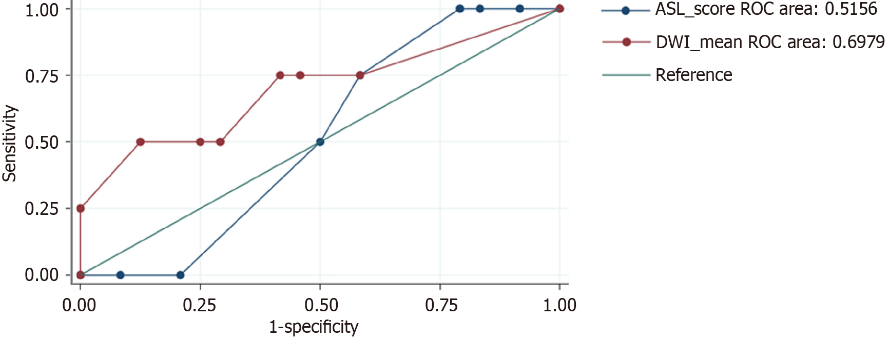
Figure 2 Relationship between arterial spin-labeling and diffusion-weighted imaging signal intensity.
There was a moderate positive relationship in the frontal lobe (0.53-0.68, P < 0.05) and a low positive correlation in the temporal, parietal, and occipital lobes (0.36-0.49, P < 0.05). Normalized data showed a low-moderate positive correlation in the frontal and occipital lobes. ROC: Receiver operator characteristic.
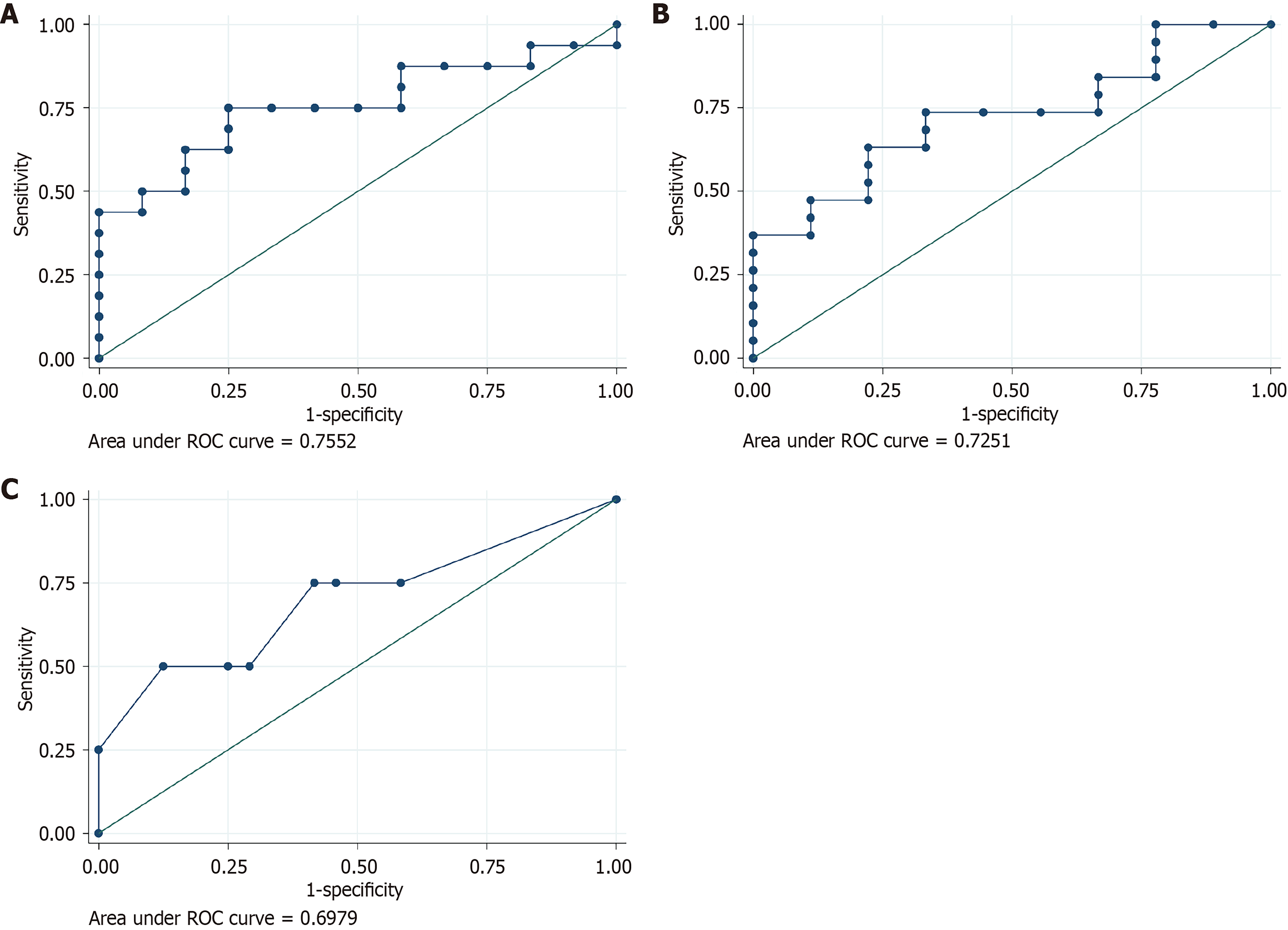
Figure 3 Relationship between arterial spin-labeling and diffusion weighted imaging signal intensity and patient outcomes.
A: Relationship between arterial spin-labeling (ASL) signal intensity within the left frontal lobe and likelihood of within one day of anoxic brain injury; B: Relationship between ASL signal intensity within the right frontal lobe and likelihood of absent brainstem reflexes (BSR) during the course of hospitalization; C: Relationship between diffusion weighted imaging signal intensity and likelihood of absent BSR during the course of hospitalization.
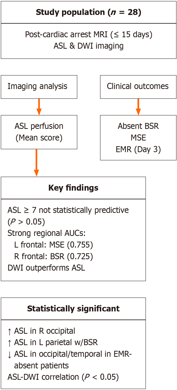
Figure 4 Visual summary of the study population, methodology, and key exploratory findings.
A total of 28 patients were included in the analysis. Clinical outcomes of interest included absent brainstem reflexes (BSR), myoclonus status epilepticus (MSE), and absent extensor or motor reflexes. While certain regional arterial spin-labeling (ASL) perfusion patterns-such as left frontal ASL signal with MSE and right frontal ASL signal with BSR-showed relatively higher area under the curve values, these associations did not reach statistical significance and should be interpreted cautiously. Diffusion-weighted imaging demonstrated relatively better predictive performance compared to ASL in this limited cohort. BSR: Brainstem reflexes; MSE: Myoclonus status epilepticus; EMR: Absent extensor or motor reflexes; ASL: Arterial spin-labeling; AUC: Area under the curve; DWI: Diffusion-weighted imaging; MRI: Magnetic resonance imaging.
- Citation: Beutler BD, Antwi-Amoabeng D, Weinert D, Shah I, Ulanja MB, Moody AE, Lei X, Lerner A, Shiroishi MS, Assadsangabi R. Prognostic value of arterial spin-labeling perfusion in anoxic brain injury: A retrospective cohort study. World J Radiol 2025; 17(8): 111065
- URL: https://www.wjgnet.com/1949-8470/full/v17/i8/111065.htm
- DOI: https://dx.doi.org/10.4329/wjr.v17.i8.111065


