Copyright
©The Author(s) 2025.
World J Radiol. Aug 28, 2025; 17(8): 111065
Published online Aug 28, 2025. doi: 10.4329/wjr.v17.i8.111065
Published online Aug 28, 2025. doi: 10.4329/wjr.v17.i8.111065
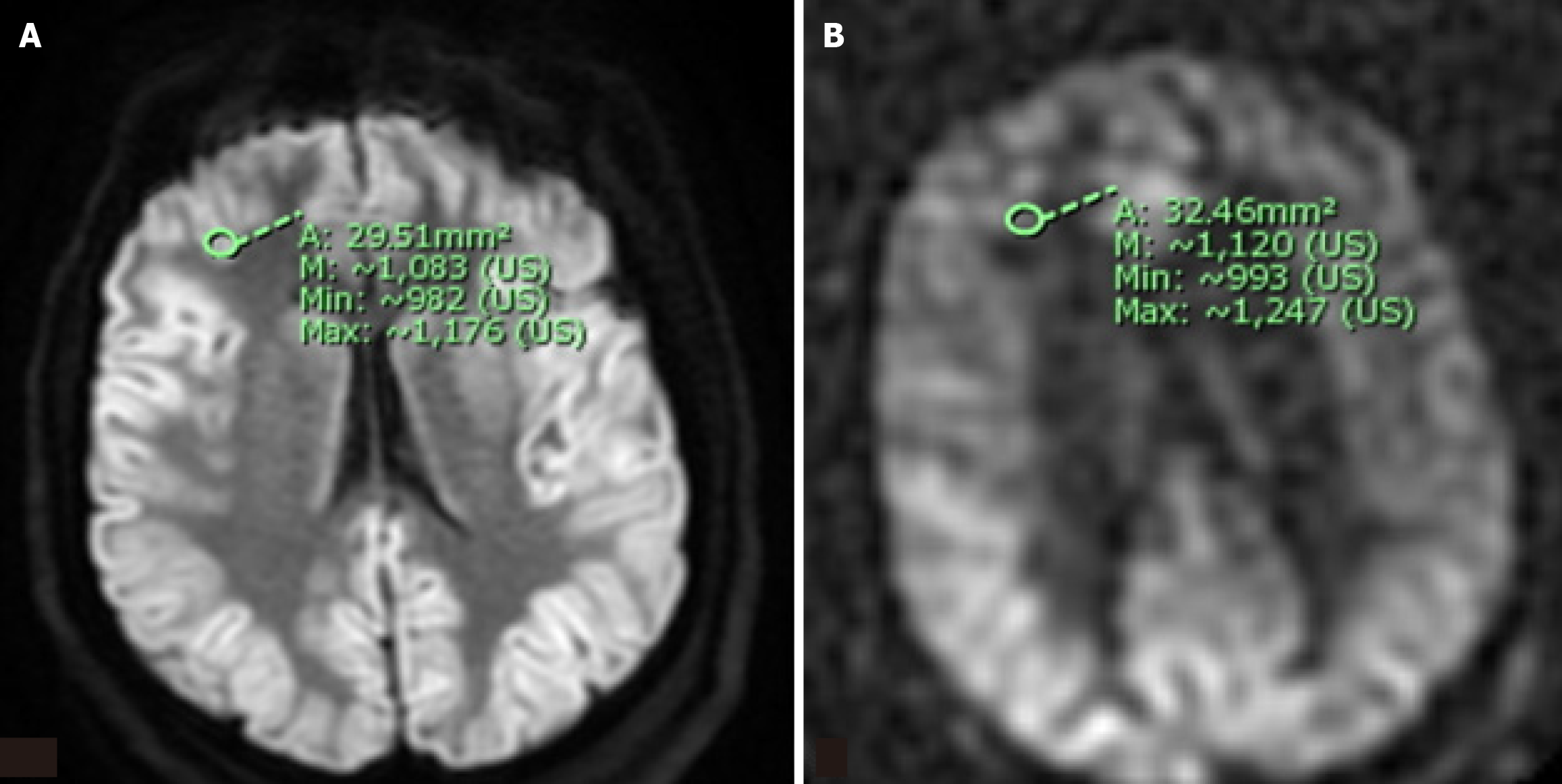
Figure 1 Representative diffusion weighted imaging and arterial spin-labeling sequences illustrating region of interest selection for semiquantiative analysis of signal intensity.
A region of interest (ROI) measuring approximately 30 mm² areas was selected for each of twelve brain regions (bilateral frontal, parietal, temporal, and occipital lobes; thalami; and basal ganglia). The raw mean and maximum signal intensity within each ROI was recorded and normalized on a 0-12 point scale using the signal intensity within the ipsilateral cerebellar hemisphere as an internal reference value. A: A representative example of an ROI within the right frontal lobe on a diffusion weighted imaging sequence; B: A representative example of an ROI within the right frontal lobe on the corresponding arterial spin-labeling sequence.
- Citation: Beutler BD, Antwi-Amoabeng D, Weinert D, Shah I, Ulanja MB, Moody AE, Lei X, Lerner A, Shiroishi MS, Assadsangabi R. Prognostic value of arterial spin-labeling perfusion in anoxic brain injury: A retrospective cohort study. World J Radiol 2025; 17(8): 111065
- URL: https://www.wjgnet.com/1949-8470/full/v17/i8/111065.htm
- DOI: https://dx.doi.org/10.4329/wjr.v17.i8.111065


