Copyright
©The Author(s) 2025.
World J Radiol. Aug 28, 2025; 17(8): 111065
Published online Aug 28, 2025. doi: 10.4329/wjr.v17.i8.111065
Published online Aug 28, 2025. doi: 10.4329/wjr.v17.i8.111065
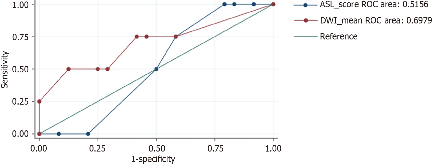
Figure 2 Relationship between arterial spin-labeling and diffusion-weighted imaging signal intensity.
There was a moderate positive relationship in the frontal lobe (0.53-0.68, P < 0.05) and a low positive correlation in the temporal, parietal, and occipital lobes (0.36-0.49, P < 0.05). Normalized data showed a low-moderate positive correlation in the frontal and occipital lobes. ROC: Receiver operator characteristic.
- Citation: Beutler BD, Antwi-Amoabeng D, Weinert D, Shah I, Ulanja MB, Moody AE, Lei X, Lerner A, Shiroishi MS, Assadsangabi R. Prognostic value of arterial spin-labeling perfusion in anoxic brain injury: A retrospective cohort study. World J Radiol 2025; 17(8): 111065
- URL: https://www.wjgnet.com/1949-8470/full/v17/i8/111065.htm
- DOI: https://dx.doi.org/10.4329/wjr.v17.i8.111065


