
















Copyright: ©Author(s) 2026.
World J Gastrointest Surg. Nov 27, 2026; 18(11): 125359
Published online Nov 27, 2026. doi: 10.4240/wjgs.125359
Published online Nov 27, 2026. doi: 10.4240/wjgs.125359
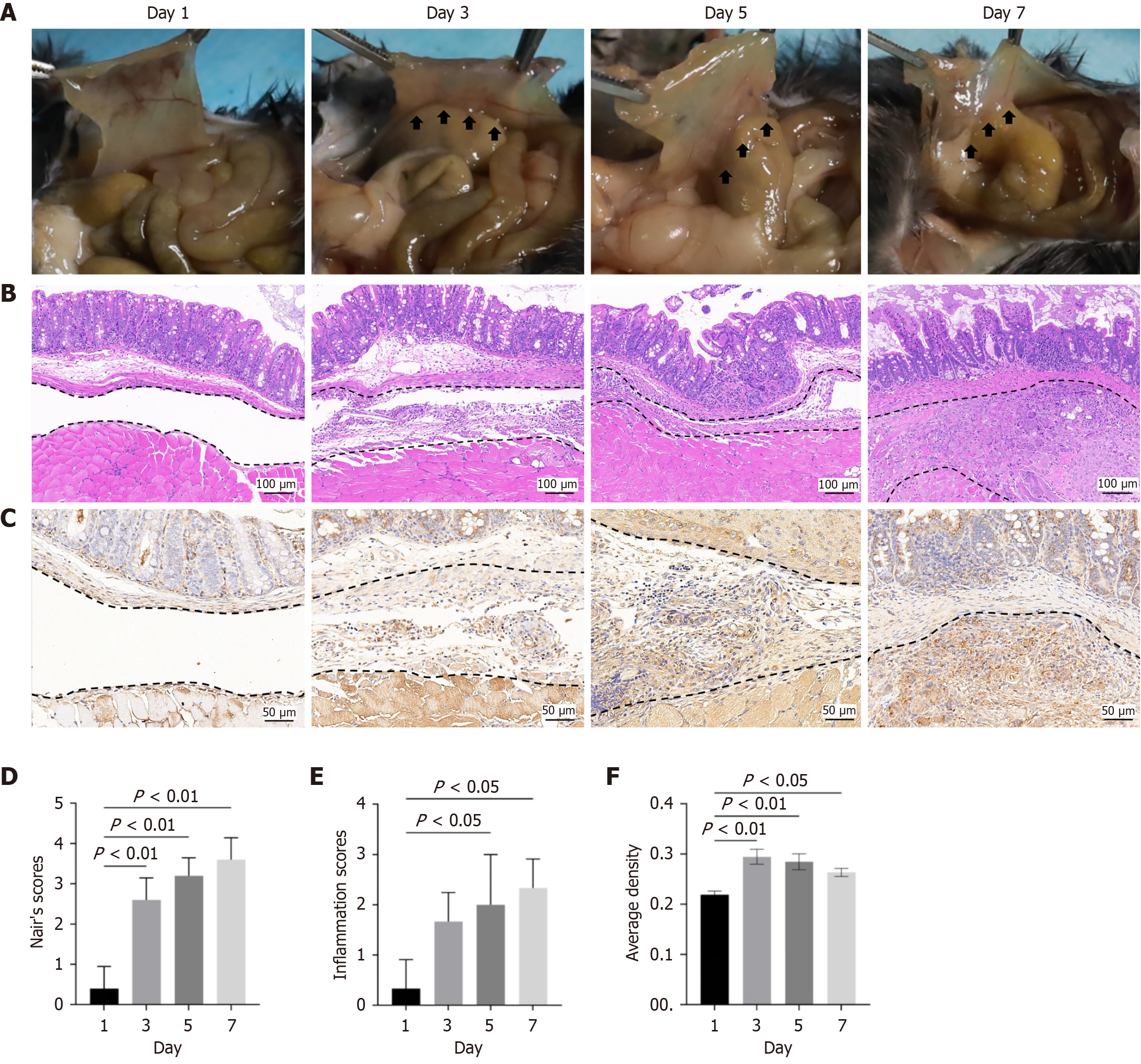
Figure 1 Thrombospondin-1 expression is increased in adhesion tissues after peritoneal injury in mice (four groups, n = 5 mice/group).
A: Representative images showing the gross postoperative abdominal adhesion conditions 1 day, 3 days, 5 days, and 7 days after peritoneal injury. The arrows indicate adhesive tissue; B: Typical hematoxylin-eosin staining images of adhesion tissues 1 day, 3 days, 5 days, and 7 days after peritoneal injury; C: Typical immuno
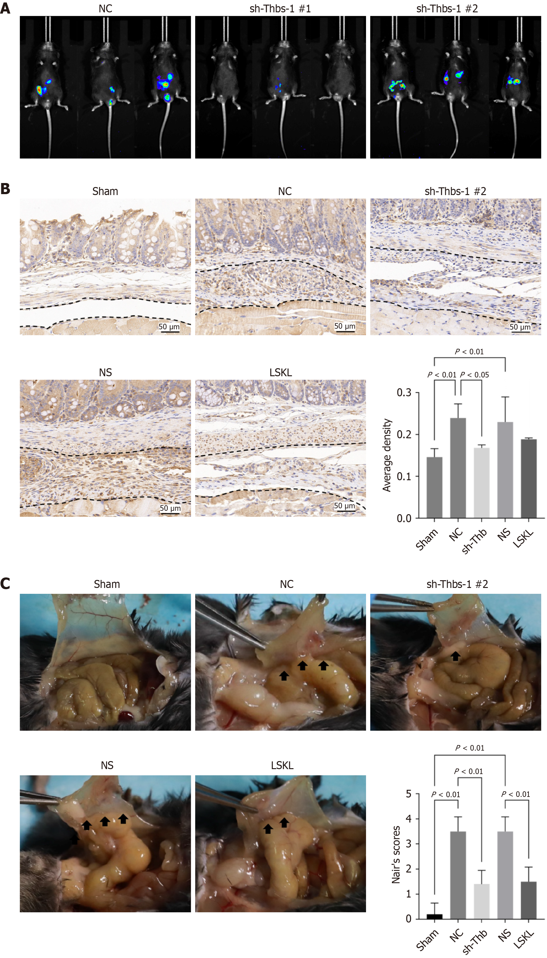
Figure 2 Thrombospondin-1 deficiency can attenuate postoperative abdominal adhesion formation in mice.
A: Typical images of live imaging for mice transfected by lentivirus (three groups, n = 3 mice/group); B: Typical immunohistochemistry staining images of thrombospondin-1 and quantification in different groups (five groups, n = 5 mice/group); C: Representative images showing the gross postoperative abdominal adhesion conditions and Nair’s score in different groups after peritoneal injury (five groups, n = 5 mice/group). The arrows indicate adhesive tissue. NC: Negative control; Thbs-1: Thrombospondin-1; NS: Normal saline; LSKL: Leu-Ser-Lys-Leu.
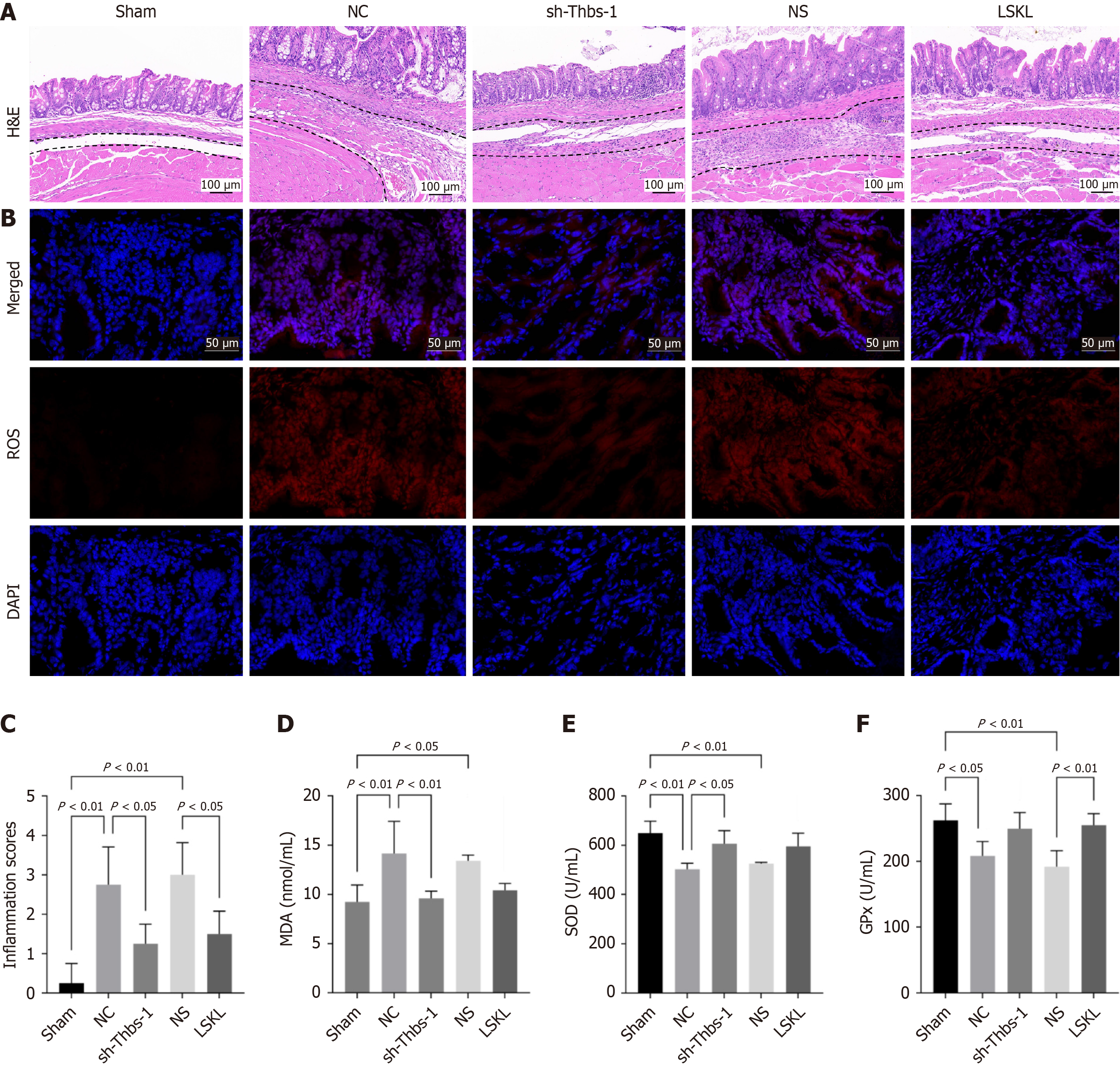
Figure 3 Thrombospondin-1 deficiency can reduce inflammation and reactive oxygen species formation in adhesion tissue (five groups, n = 5 mice/group).
A: Typical hematoxylin-eosin staining images of adhesion tissues in different groups after peritoneal injury; B: Representative images showing reactive oxygen species accumulation (red fluorescence), DAPI (blue fluorescence) and merge in adhesion tissues after peritoneal injury; C: Inflammation score in different groups; D: Malondialdehyde levels were evaluated in adhesion tissues; E: Superoxide dismutase levels were evaluated in adhesion tissues; F: Glutathione peroxidase levels were evaluated in adhesion tissues. H&E: Hematoxylin-eosin; NC: Negative control; Thbs-1: Thrombospondin-1; NS: Normal saline; LSKL: Leu-Ser-Lys-Leu; ROS: Reactive oxygen species; MDA: Malondialdehyde; SOD: Superoxide dismutase; GPx: Glutathione peroxidase.
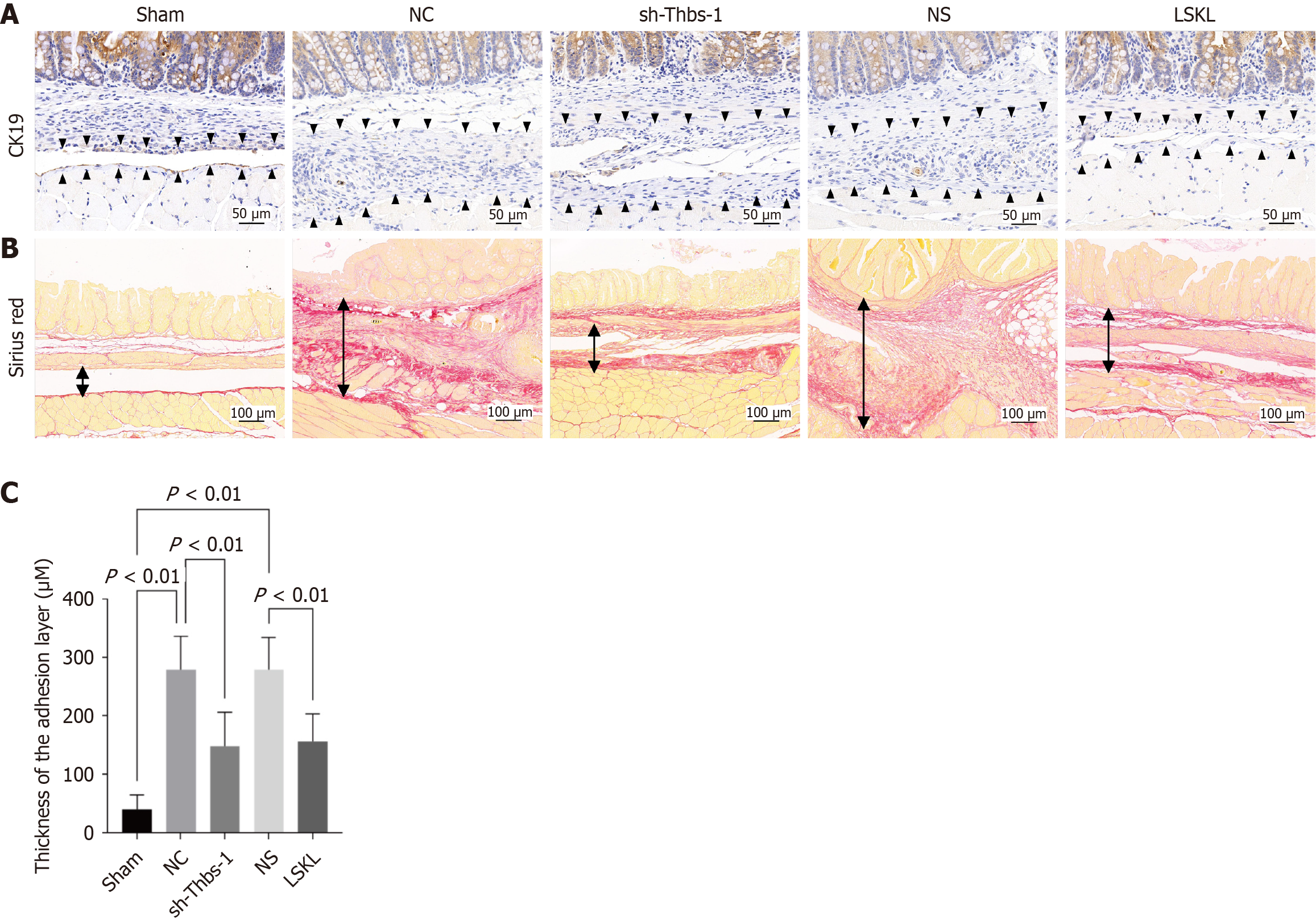
Figure 4 Thrombospondin-1 deficiency promotes peritoneal mesothelial cell repair (five groups, n = 5 mice/group).
A: Typical picture of CK19 immunohistochemistry staining in different groups. The black arrow indicates the adhesion tissue or normal peritoneal tissues; B: Typical images of Sirius red staining in different groups, the area between the black arrows indicates adhesive tissue; C: Fiber thickness in different groups as assessed by Sirius red staining. NC: Negative control; Thbs-1: Thrombospondin-1; NS: Normal saline; LSKL: Leu-Ser-Lys-Leu.
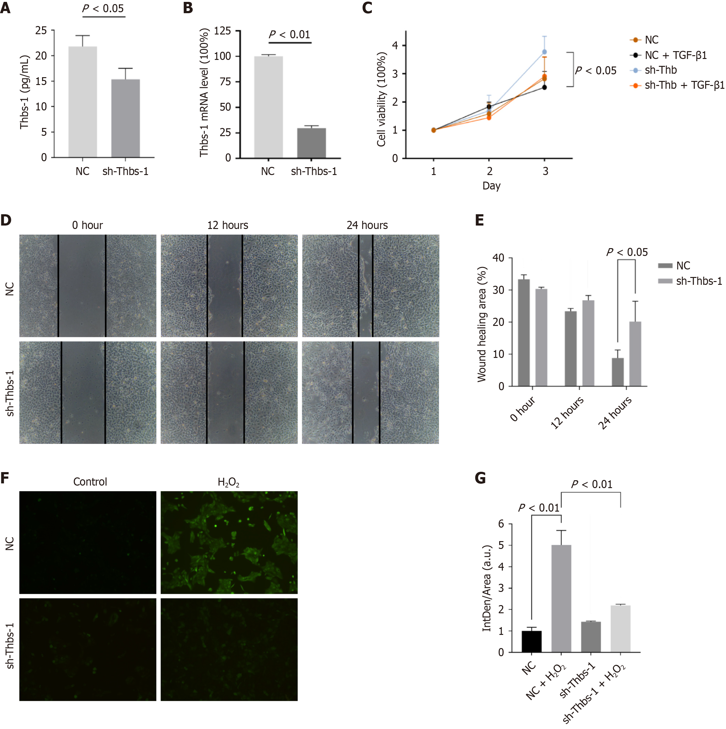
Figure 5 Thrombospondin-1 deficiency is a protective factor in mesothelial cell line.
A: The levels of thrombospondin-1 (Thbs-1) protein in the supernatant of the HMrSV5 cell culture medium were measured by enzyme-linked immunosorbent assay. n = 3; B: Quantitative real-time polymerase chain reaction was used to assess Thbs-1 mRNA expression in different groups. n = 5; C: The cell viability of HMrSV5 cells after transforming growth factor-β exposure measured by Cell Counting Kit-8 assay; D: Images of scratch-wound assay in different groups at 0 hour, 12 hours and 24 hours’ time intervals post injury. n = 5, 100 × magnification; E: Cell migration ratio in different groups. n = 5; F: Reduced reactive oxygen species (ROS) levels of HMrSV5 cells treated with H2O2 for 1 hour observed by fluorescence microscopy. n = 3, 200 × magnification; G: Quantification of the ROS levels of mesothelial cells observed by fluorescence microscopy. NC: Negative control; Thbs-1: Thrombospondin-1.
- Citation: Tong HC, Ye CC, Zhang ZX, Lin WH, Qu C, Gao YY, Wang JH, Wu YH, Liu YT. Silencing of thrombospondin-1 prevents abdominal adhesion formation by reducing inflammation and oxidative stress. World J Gastrointest Surg 2026; 18(11): 125359
- URL: https://www.wjgnet.com/1948-9366/full/v18/i11/125359.htm
- DOI: https://dx.doi.org/10.4240/wjgs.125359