
















Copyright: ©Author(s) 2026.
World J Gastrointest Surg. Nov 27, 2026; 18(11): 125359
Published online Nov 27, 2026. doi: 10.4240/wjgs.125359
Published online Nov 27, 2026. doi: 10.4240/wjgs.125359
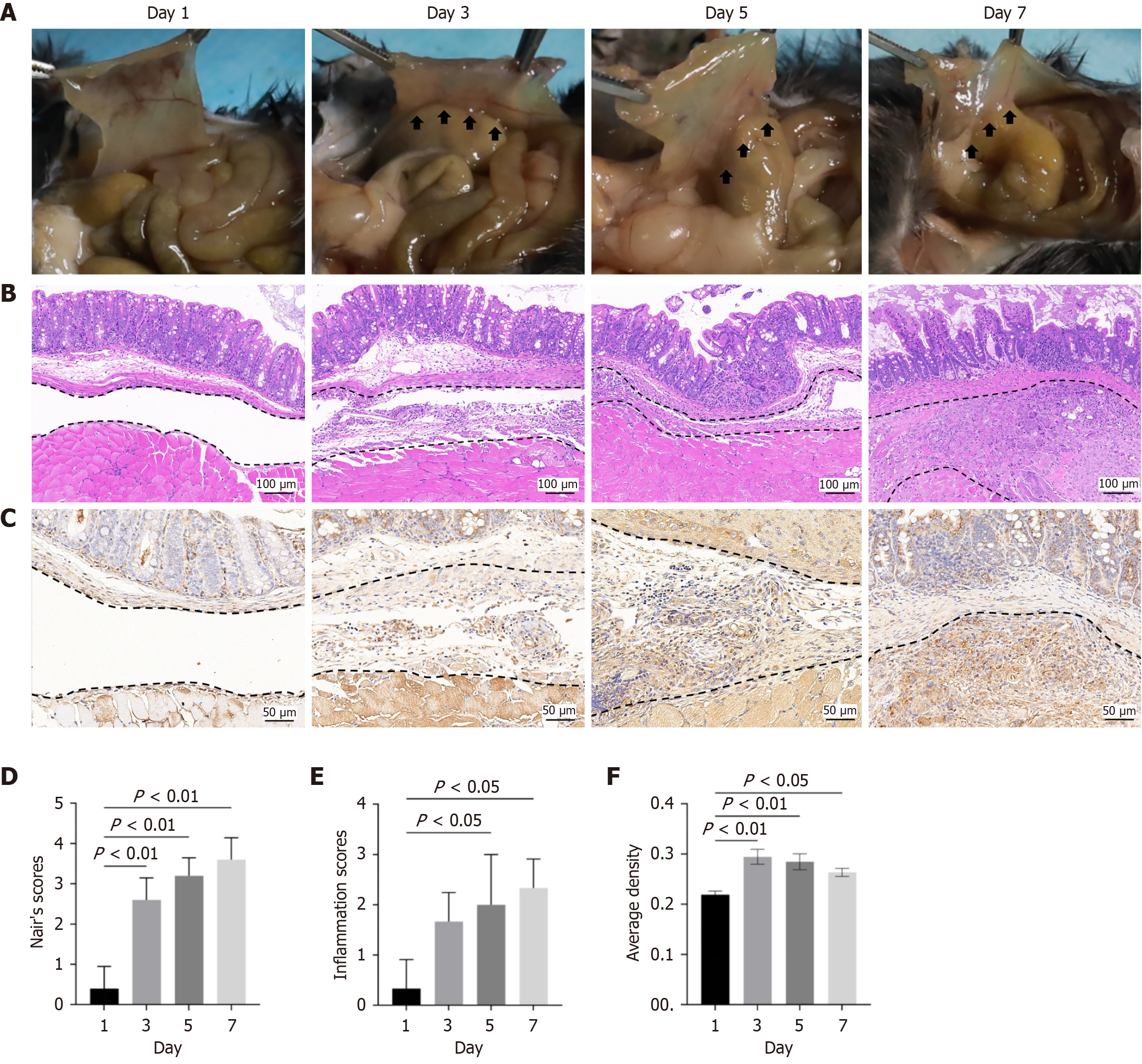
Figure 1 Thrombospondin-1 expression is increased in adhesion tissues after peritoneal injury in mice (four groups, n = 5 mice/group).
A: Representative images showing the gross postoperative abdominal adhesion conditions 1 day, 3 days, 5 days, and 7 days after peritoneal injury. The arrows indicate adhesive tissue; B: Typical hematoxylin-eosin staining images of adhesion tissues 1 day, 3 days, 5 days, and 7 days after peritoneal injury; C: Typical immuno
- Citation: Tong HC, Ye CC, Zhang ZX, Lin WH, Qu C, Gao YY, Wang JH, Wu YH, Liu YT. Silencing of thrombospondin-1 prevents abdominal adhesion formation by reducing inflammation and oxidative stress. World J Gastrointest Surg 2026; 18(11): 125359
- URL: https://www.wjgnet.com/1948-9366/full/v18/i11/125359.htm
- DOI: https://dx.doi.org/10.4240/wjgs.125359