
















Copyright: ©Author(s) 2026.
World J Gastrointest Surg. Nov 27, 2026; 18(11): 124761
Published online Nov 27, 2026. doi: 10.4240/wjgs.124761
Published online Nov 27, 2026. doi: 10.4240/wjgs.124761

Figure 1 Timeline of the patient’s medical visits over a 10-year period.
RCC: Renal cell carcinoma; PET-CT: Positron emission tomography - computed tomography; MDT: Multidisciplinary team; ccRCC: Clear cell renal cell carcinoma.
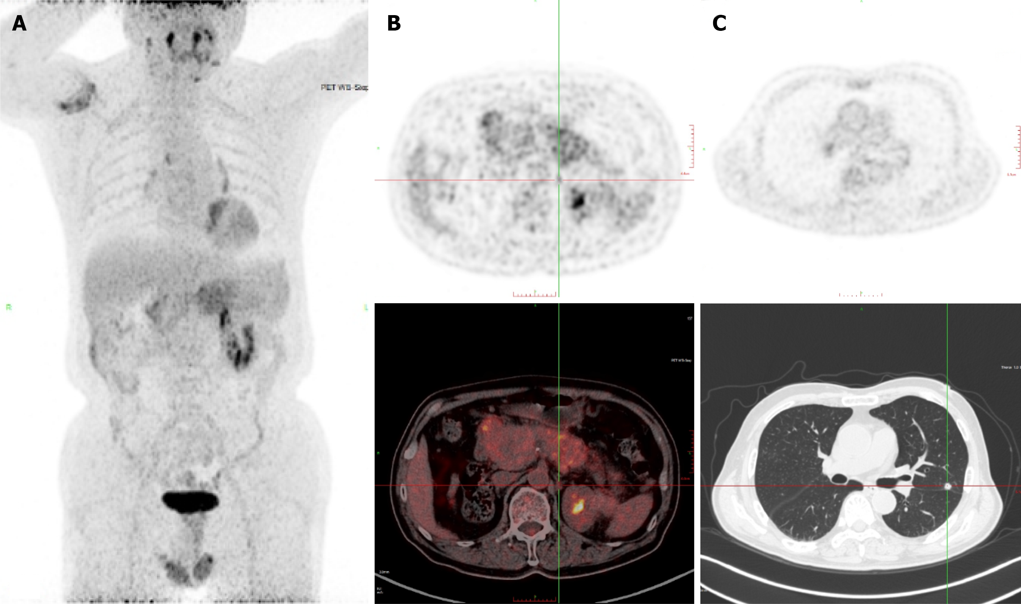
Figure 2 Recurrent and metastatic lesions in the lungs, left renal pelvis, and retroperitoneal soft tissues were identified via positron emission tomography/computed tomography in December 2022.
A: Hyperdense nodule with slightly increased 18F-fluorodeoxyglucose (18F-FDG) uptake in the left renal pelvis; B: Soft tissue mass with increased 18F-FDG uptake in the space between the upper pole of the left kidney and the posterior aspect of the pancreatic tail; C: Round solid nodule with slightly increased 18F-FDG uptake was detected in the left lung, suggestive of pulmonary metastatic tumors.
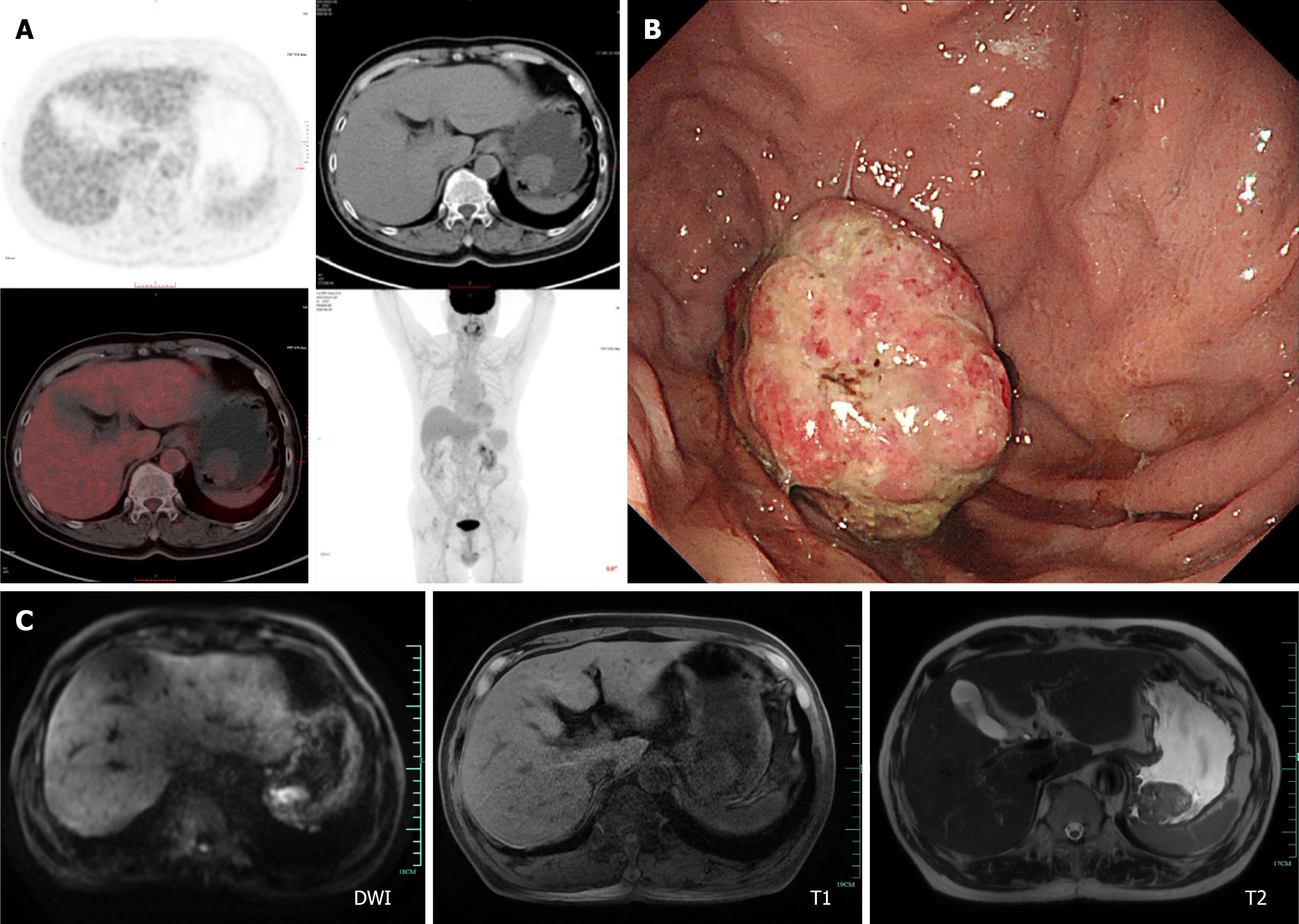
Figure 3 Gastric metastases were detected by positron emission tomography/computed tomography in June 2025, followed by endoscopy.
A: Soft tissue density mass with increased 18F-fluorodeoxyglucose uptake was identified in the gastric fundus, representing a new lesion compared with prior imaging studies; B: Spherical mass in the gastric fundus with superficial erosion; C: Diffusion-weighted imaging, T1-weighted and T2-weighted magnetic resonance images of the gastric fundus lesion. DWI: Diffusion weighted imaging.
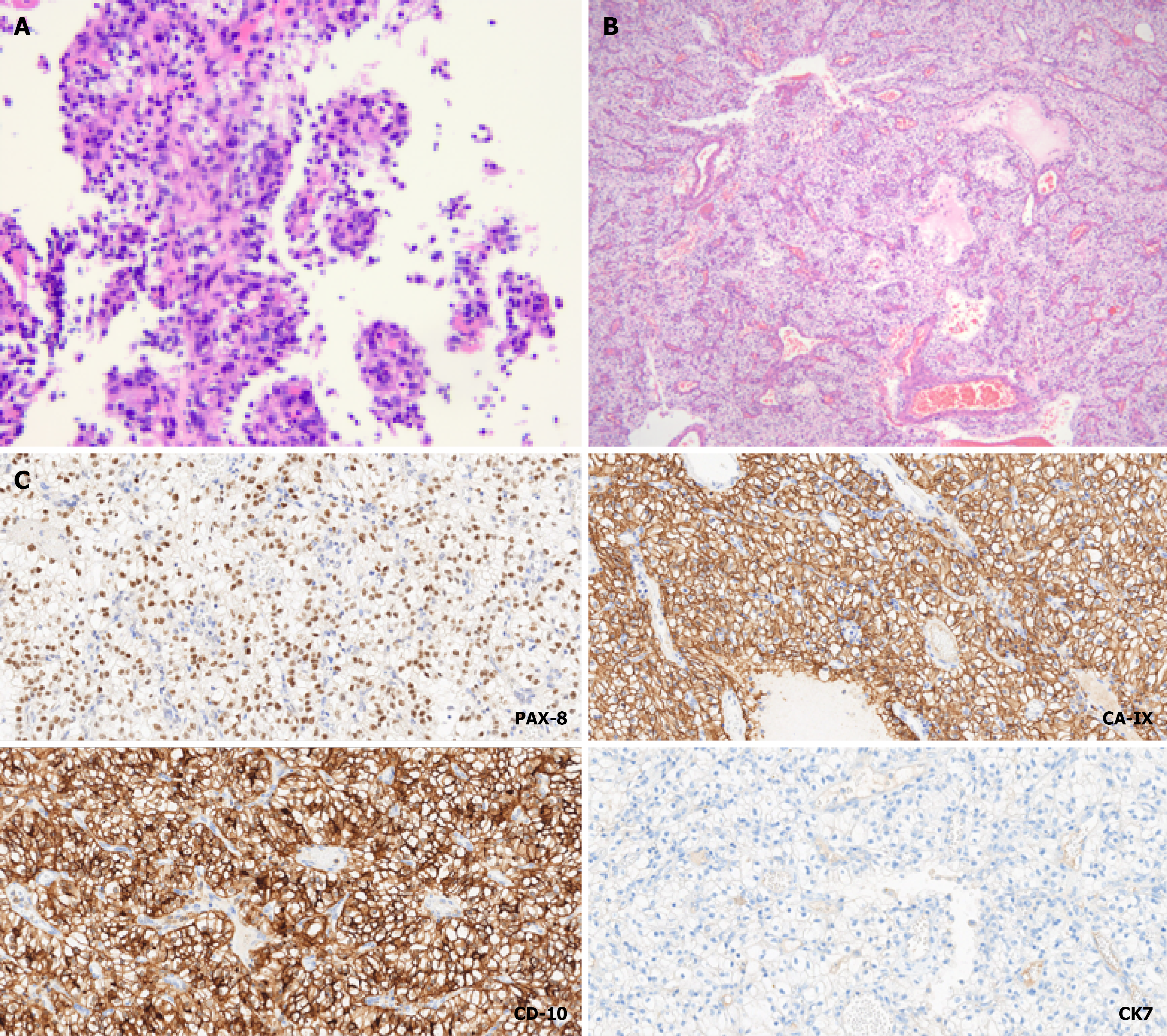
Figure 4 Diagnosis of gastric metastasis from clear cell renal cell carcinoma was confirmed via pathological immunohistochemical analysis of the lesion.
A: Microscopic images of pathological sections obtained via endoscopic gastric biopsy [hematoxylin and eosin (H&E), 100 ×]; B: Microscopic images of sections from the gross surgical resection specimen (H&E, 40 ×); C: Postoperative pathological immunohistochemical staining results revealed positivity for PAX-8 (200 ×), carbonic anhydrase IX (200 ×), and CD10 (200 ×), and negativity for cytokeratin 7 (200 ×), which was consistent with the diagnosis of clear cell renal cell carcinoma.
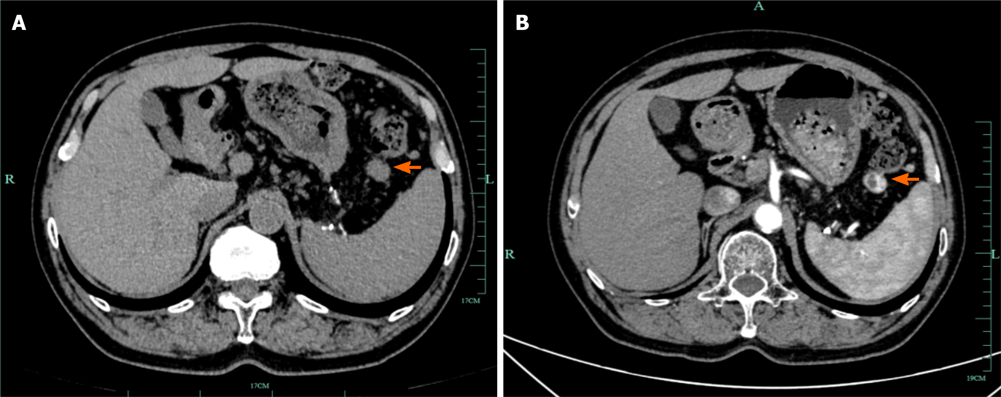
Figure 5 Postoperative surveillance computed tomography.
A: Non-contrast computed tomography (CT) at 4 months after surgery (December 2025) showing expected post-gastrectomy changes with minimal residual exudate at the resection bed. A newly identified in the left mesentery (arrow) warrants contrast-enhanced CT evaluation. No other sites of disease progression are evident; B: Contrast-enhanced CT at 10 months after surgery (June 2026) demonstrating the left mesenteric nodule (arrow) to be stable in size and showing heterogeneous enhancement compared with the December 2025 examination, without evidence of radiologic progression. No local recurrence is observed at the gastric resection bed.
- Citation: Gao TZ, Zheng CZ, Zhu KK, Wu XN, He CY, Lou YM, Liu XS. Gastric metastasis from renal cell carcinoma with a 10-year interval: A case report. World J Gastrointest Surg 2026; 18(11): 124761
- URL: https://www.wjgnet.com/1948-9366/full/v18/i11/124761.htm
- DOI: https://dx.doi.org/10.4240/wjgs.124761