
















Copyright: ©Author(s) 2026.
World J Gastrointest Surg. Nov 27, 2026; 18(11): 124761
Published online Nov 27, 2026. doi: 10.4240/wjgs.124761
Published online Nov 27, 2026. doi: 10.4240/wjgs.124761
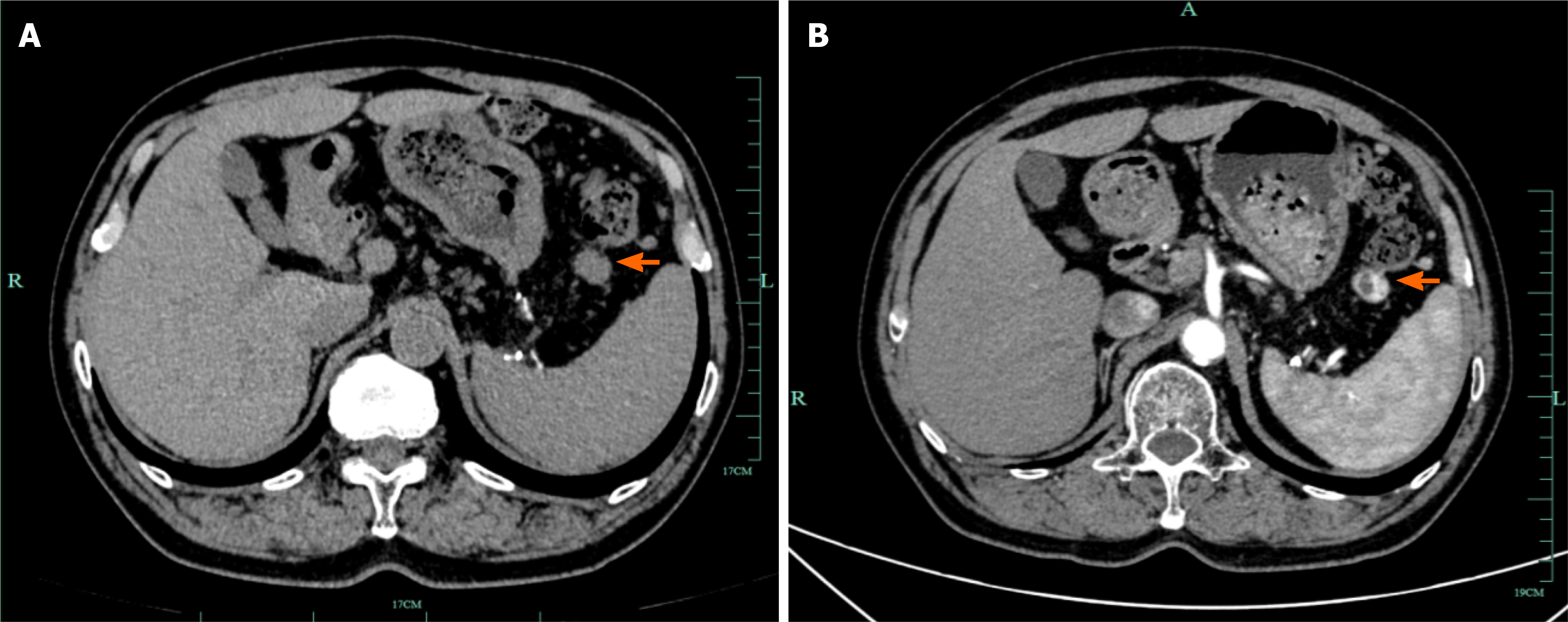
Figure 5 Postoperative surveillance computed tomography.
A: Non-contrast computed tomography (CT) at 4 months after surgery (December 2025) showing expected post-gastrectomy changes with minimal residual exudate at the resection bed. A newly identified in the left mesentery (arrow) warrants contrast-enhanced CT evaluation. No other sites of disease progression are evident; B: Contrast-enhanced CT at 10 months after surgery (June 2026) demonstrating the left mesenteric nodule (arrow) to be stable in size and showing heterogeneous enhancement compared with the December 2025 examination, without evidence of radiologic progression. No local recurrence is observed at the gastric resection bed.
- Citation: Gao TZ, Zheng CZ, Zhu KK, Wu XN, He CY, Lou YM, Liu XS. Gastric metastasis from renal cell carcinoma with a 10-year interval: A case report. World J Gastrointest Surg 2026; 18(11): 124761
- URL: https://www.wjgnet.com/1948-9366/full/v18/i11/124761.htm
- DOI: https://dx.doi.org/10.4240/wjgs.124761