
















Copyright: ©Author(s) 2026.
World J Gastrointest Surg. Nov 27, 2026; 18(11): 124761
Published online Nov 27, 2026. doi: 10.4240/wjgs.124761
Published online Nov 27, 2026. doi: 10.4240/wjgs.124761
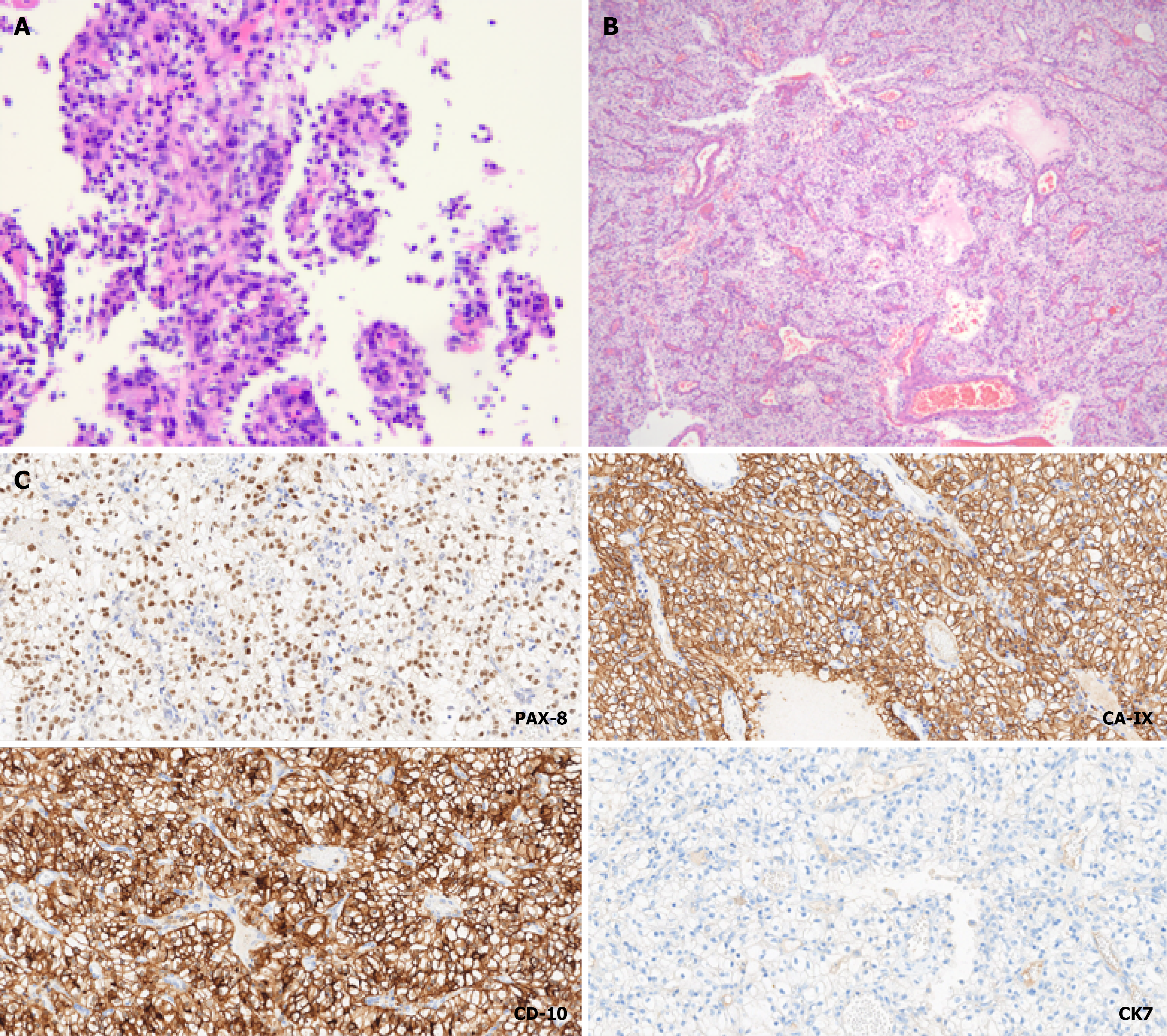
Figure 4 Diagnosis of gastric metastasis from clear cell renal cell carcinoma was confirmed via pathological immunohistochemical analysis of the lesion.
A: Microscopic images of pathological sections obtained via endoscopic gastric biopsy [hematoxylin and eosin (H&E), 100 ×]; B: Microscopic images of sections from the gross surgical resection specimen (H&E, 40 ×); C: Postoperative pathological immunohistochemical staining results revealed positivity for PAX-8 (200 ×), carbonic anhydrase IX (200 ×), and CD10 (200 ×), and negativity for cytokeratin 7 (200 ×), which was consistent with the diagnosis of clear cell renal cell carcinoma.
- Citation: Gao TZ, Zheng CZ, Zhu KK, Wu XN, He CY, Lou YM, Liu XS. Gastric metastasis from renal cell carcinoma with a 10-year interval: A case report. World J Gastrointest Surg 2026; 18(11): 124761
- URL: https://www.wjgnet.com/1948-9366/full/v18/i11/124761.htm
- DOI: https://dx.doi.org/10.4240/wjgs.124761