
















Copyright: ©Author(s) 2026.
World J Gastrointest Surg. Nov 27, 2026; 18(11): 124761
Published online Nov 27, 2026. doi: 10.4240/wjgs.124761
Published online Nov 27, 2026. doi: 10.4240/wjgs.124761
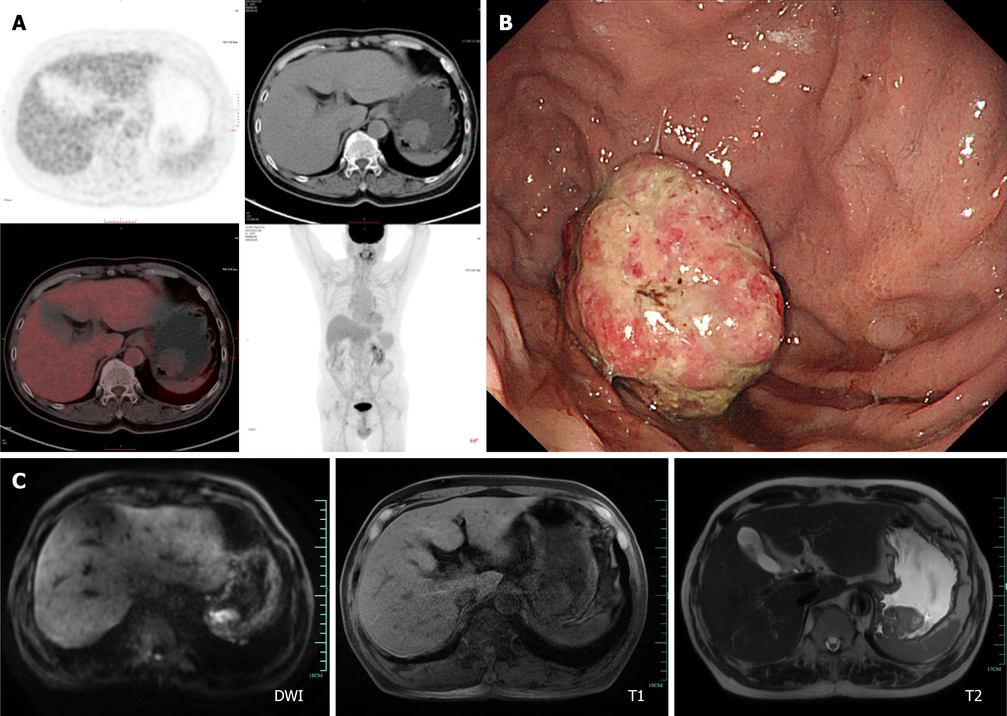
Figure 3 Gastric metastases were detected by positron emission tomography/computed tomography in June 2025, followed by endoscopy.
A: Soft tissue density mass with increased 18F-fluorodeoxyglucose uptake was identified in the gastric fundus, representing a new lesion compared with prior imaging studies; B: Spherical mass in the gastric fundus with superficial erosion; C: Diffusion-weighted imaging, T1-weighted and T2-weighted magnetic resonance images of the gastric fundus lesion. DWI: Diffusion weighted imaging.
- Citation: Gao TZ, Zheng CZ, Zhu KK, Wu XN, He CY, Lou YM, Liu XS. Gastric metastasis from renal cell carcinoma with a 10-year interval: A case report. World J Gastrointest Surg 2026; 18(11): 124761
- URL: https://www.wjgnet.com/1948-9366/full/v18/i11/124761.htm
- DOI: https://dx.doi.org/10.4240/wjgs.124761