Copyright
©The Author(s) 2025.
World J Gastrointest Oncol. May 15, 2025; 17(5): 104802
Published online May 15, 2025. doi: 10.4251/wjgo.v17.i5.104802
Published online May 15, 2025. doi: 10.4251/wjgo.v17.i5.104802
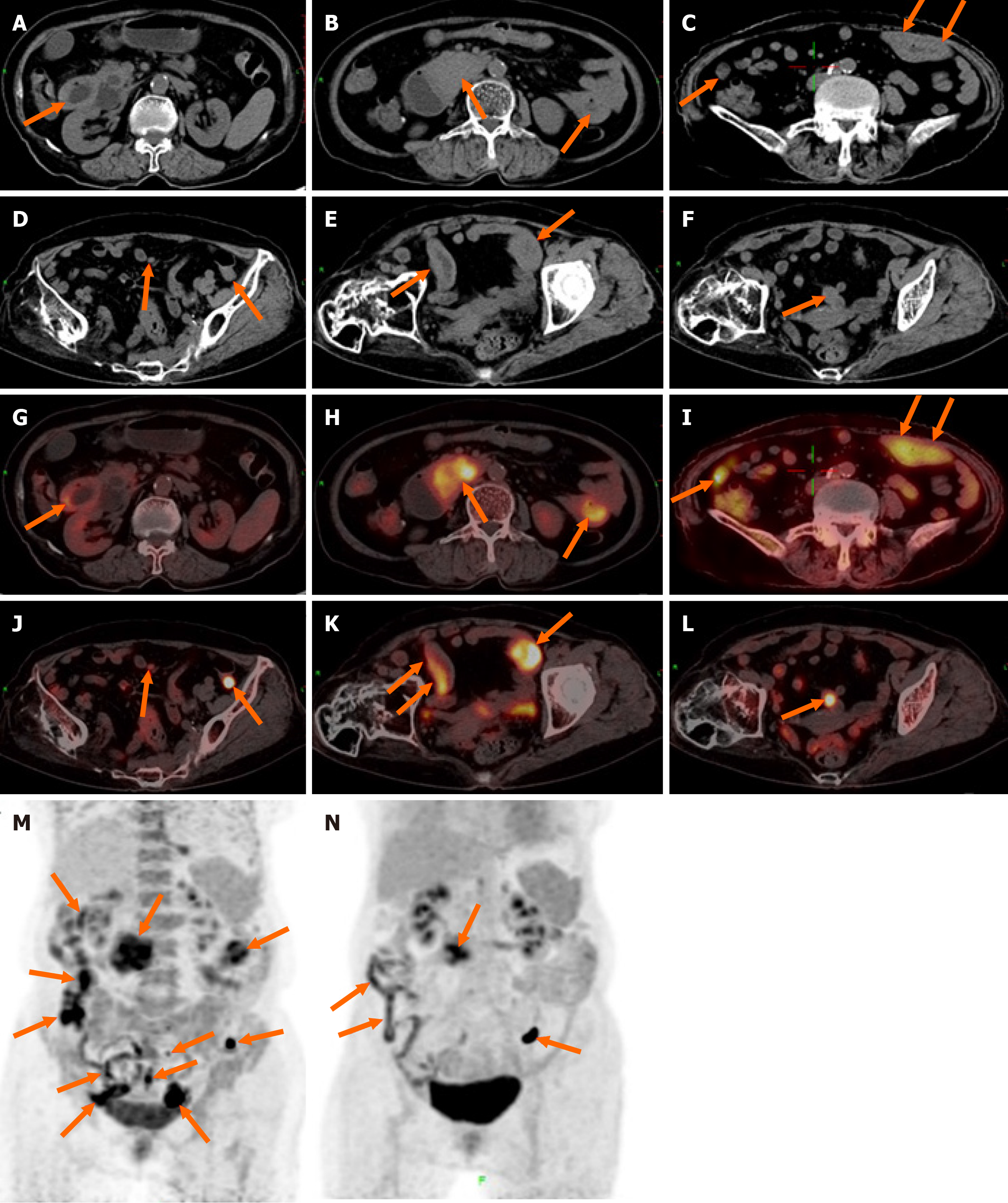
Figure 1 18F-fluorodeoxyglucose positron emission tomography/computed tomography findings of monomorphic epitheliotropic intestinal T-cell lymphoma.
A-L: Multiple segmental thickenings were observed in the descending and horizontal segments of the duodenum, jejunum, and ileum, accompanied by enlarged lymph nodes scattered throughout the abdominal and pelvic mesentery; M: Functional images of the abdomen at the positron emission tomography level before treatment; N: After completing three cycles of chemotherapy, the patient underwent a follow-up 18F-fluorodeoxyglucose positron emission tomography/computed tomography scan, which revealed a decrease in multiple lesions in the small intestine as well as a reduction in enlarged mesenteric lymph nodes.
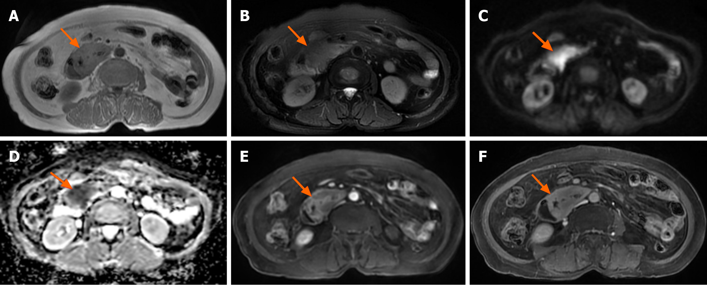
Figure 2 Magnetic resonance imaging findings of monomorphic epitheliotropic intestinal T-cell lymphoma.
A: The lesion in the horizontal segment of the duodenum showed an iso-signal on T1 weighted imaging; B: Iso- or hypo-signals on T2 weighted imaging; C: Limited diffusion on diffusion-weighted imaging; D: Apparent diffusion coefficient value of 0.8 × 10⁻³ mm2/second; E and F: Delayed enhancement on an enhanced scan.
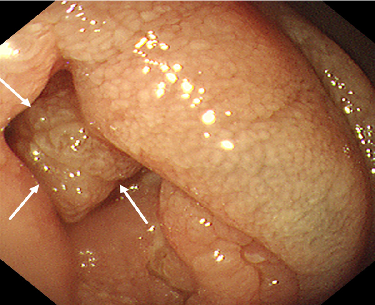
Figure 3 Gastroduodenoscopy of monomorphic epitheliotropic intestinal T-cell lymphoma.
A cauliflower-like mass was found in the duodenal lumen, resulting in significant stenosis and obstruction of the intestinal lumen.
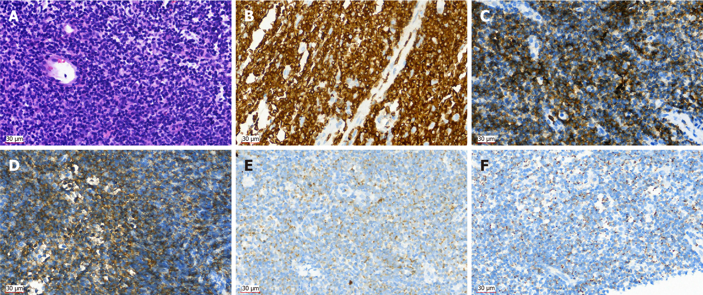
Figure 4 Histopathological and immunohistochemical examination of monomorphic epitheliotropic intestinal T-cell lymphoma.
A: Atypical lymphoid cells were diffusely proliferated, with round or irregular nuclei, thick chromatin, and scattered plasma cells (hematoxylin eosin × 40); B: Positive in CD3 (× 40); C: Positive in CD8 (× 40); D: Positive in CD56 (× 40); E: Positive in granzyme B (× 40); F: Positive in T-cell intracellular antigen 1 (× 40).
- Citation: Tang WJ, Luo SH, Wu Z, Kang Y, Lan B, Zhang ZQ, Zhong JY, Zhong JP, Wen CJ. Imaging findings of primary monomorphic epitheliotropic intestinal T-cell lymphoma: A case report. World J Gastrointest Oncol 2025; 17(5): 104802
- URL: https://www.wjgnet.com/1948-5204/full/v17/i5/104802.htm
- DOI: https://dx.doi.org/10.4251/wjgo.v17.i5.104802


