Copyright
©The Author(s) 2025.
World J Gastrointest Oncol. May 15, 2025; 17(5): 104802
Published online May 15, 2025. doi: 10.4251/wjgo.v17.i5.104802
Published online May 15, 2025. doi: 10.4251/wjgo.v17.i5.104802
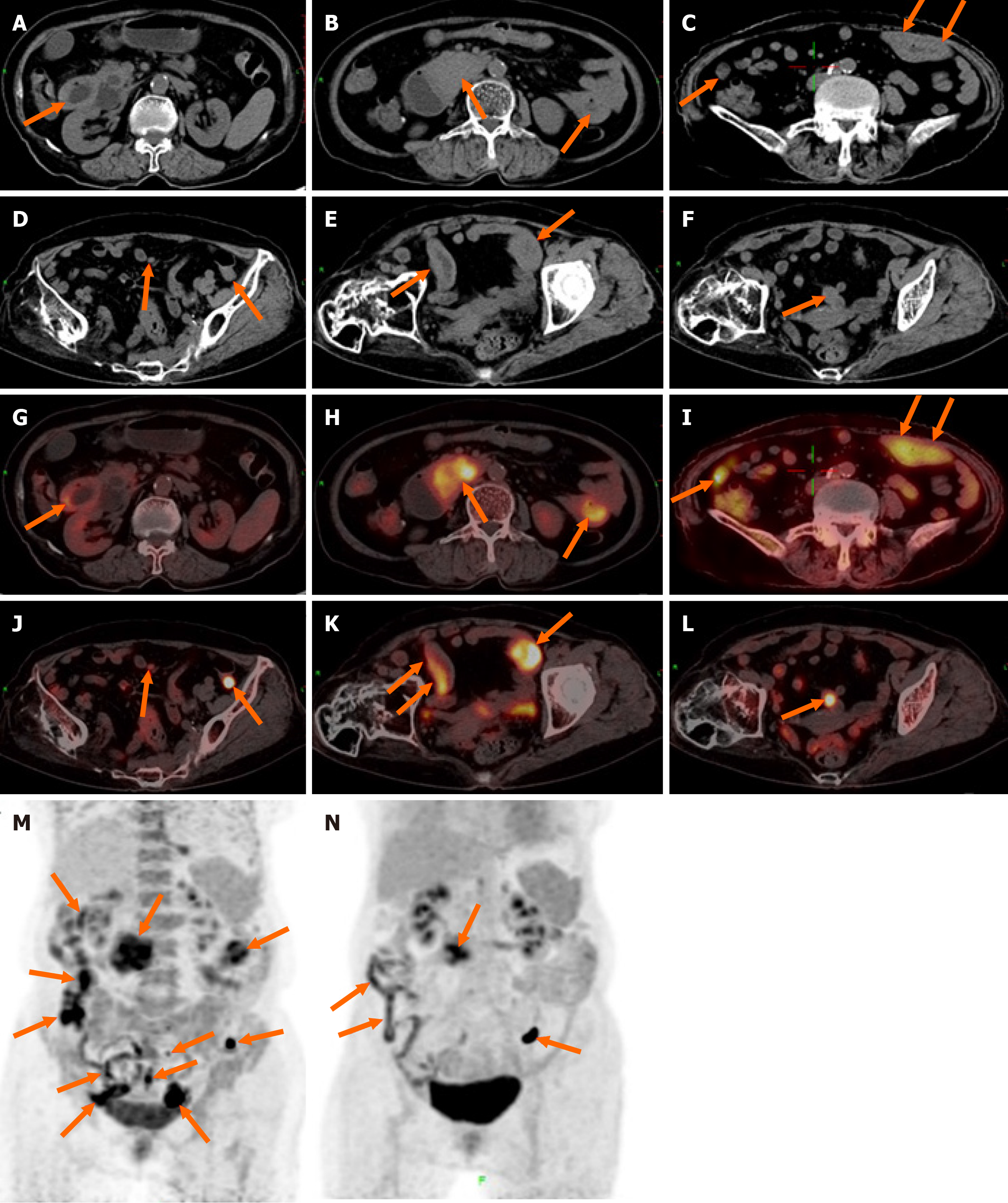
Figure 1 18F-fluorodeoxyglucose positron emission tomography/computed tomography findings of monomorphic epitheliotropic intestinal T-cell lymphoma.
A-L: Multiple segmental thickenings were observed in the descending and horizontal segments of the duodenum, jejunum, and ileum, accompanied by enlarged lymph nodes scattered throughout the abdominal and pelvic mesentery; M: Functional images of the abdomen at the positron emission tomography level before treatment; N: After completing three cycles of chemotherapy, the patient underwent a follow-up 18F-fluorodeoxyglucose positron emission tomography/computed tomography scan, which revealed a decrease in multiple lesions in the small intestine as well as a reduction in enlarged mesenteric lymph nodes.
- Citation: Tang WJ, Luo SH, Wu Z, Kang Y, Lan B, Zhang ZQ, Zhong JY, Zhong JP, Wen CJ. Imaging findings of primary monomorphic epitheliotropic intestinal T-cell lymphoma: A case report. World J Gastrointest Oncol 2025; 17(5): 104802
- URL: https://www.wjgnet.com/1948-5204/full/v17/i5/104802.htm
- DOI: https://dx.doi.org/10.4251/wjgo.v17.i5.104802


