Copyright
©The Author(s) 2025.
World J Gastrointest Oncol. May 15, 2025; 17(5): 104802
Published online May 15, 2025. doi: 10.4251/wjgo.v17.i5.104802
Published online May 15, 2025. doi: 10.4251/wjgo.v17.i5.104802
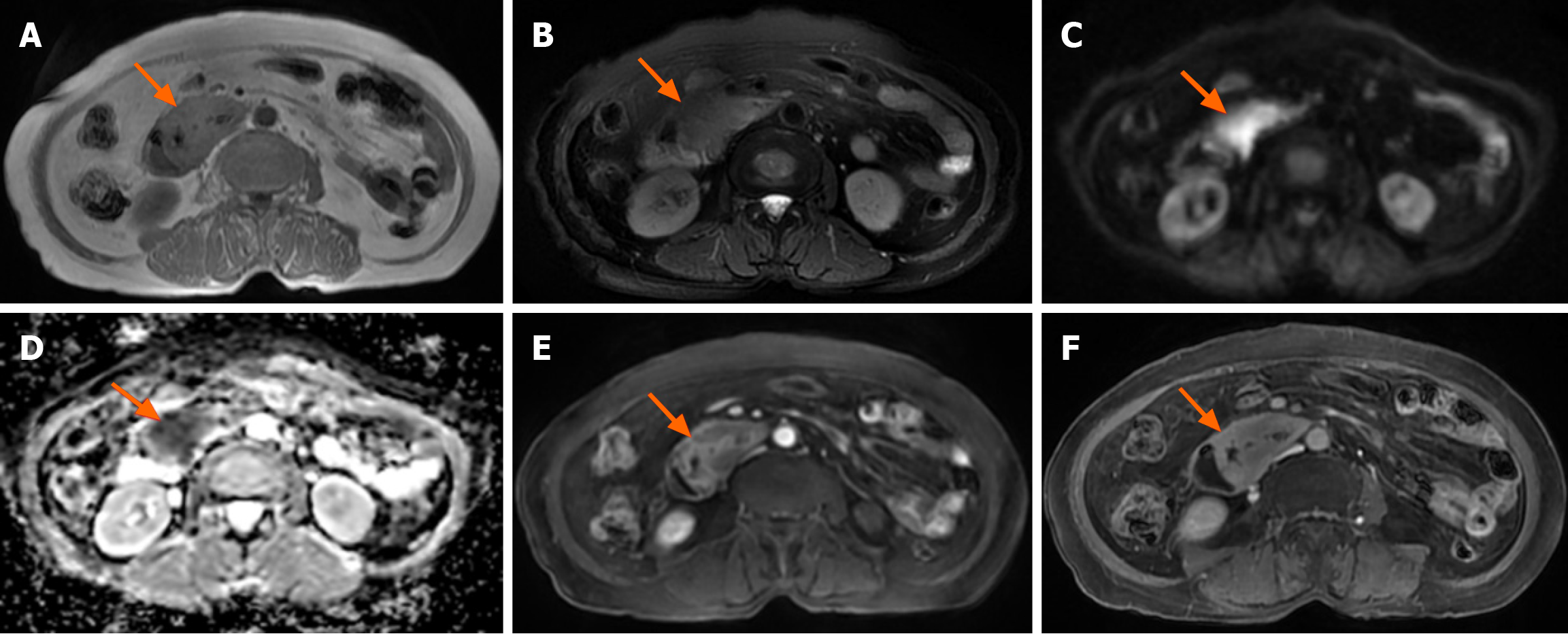
Figure 2 Magnetic resonance imaging findings of monomorphic epitheliotropic intestinal T-cell lymphoma.
A: The lesion in the horizontal segment of the duodenum showed an iso-signal on T1 weighted imaging; B: Iso- or hypo-signals on T2 weighted imaging; C: Limited diffusion on diffusion-weighted imaging; D: Apparent diffusion coefficient value of 0.8 × 10⁻³ mm2/second; E and F: Delayed enhancement on an enhanced scan.
- Citation: Tang WJ, Luo SH, Wu Z, Kang Y, Lan B, Zhang ZQ, Zhong JY, Zhong JP, Wen CJ. Imaging findings of primary monomorphic epitheliotropic intestinal T-cell lymphoma: A case report. World J Gastrointest Oncol 2025; 17(5): 104802
- URL: https://www.wjgnet.com/1948-5204/full/v17/i5/104802.htm
- DOI: https://dx.doi.org/10.4251/wjgo.v17.i5.104802


