Copyright
©The Author(s) 2025.
World J Gastrointest Oncol. May 15, 2025; 17(5): 104802
Published online May 15, 2025. doi: 10.4251/wjgo.v17.i5.104802
Published online May 15, 2025. doi: 10.4251/wjgo.v17.i5.104802
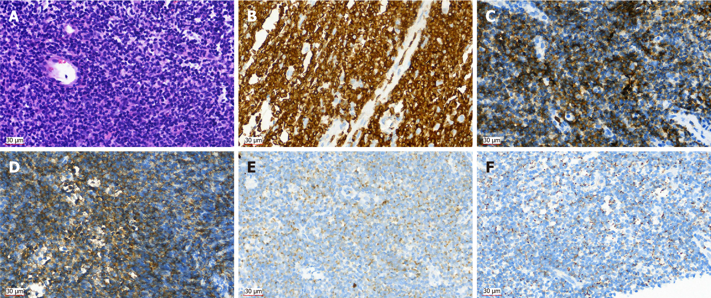
Figure 4 Histopathological and immunohistochemical examination of monomorphic epitheliotropic intestinal T-cell lymphoma.
A: Atypical lymphoid cells were diffusely proliferated, with round or irregular nuclei, thick chromatin, and scattered plasma cells (hematoxylin eosin × 40); B: Positive in CD3 (× 40); C: Positive in CD8 (× 40); D: Positive in CD56 (× 40); E: Positive in granzyme B (× 40); F: Positive in T-cell intracellular antigen 1 (× 40).
- Citation: Tang WJ, Luo SH, Wu Z, Kang Y, Lan B, Zhang ZQ, Zhong JY, Zhong JP, Wen CJ. Imaging findings of primary monomorphic epitheliotropic intestinal T-cell lymphoma: A case report. World J Gastrointest Oncol 2025; 17(5): 104802
- URL: https://www.wjgnet.com/1948-5204/full/v17/i5/104802.htm
- DOI: https://dx.doi.org/10.4251/wjgo.v17.i5.104802


