
















Copyright: ©Author(s) 2026.
World J Gastrointest Endosc. Oct 16, 2026; 18(10): 126399
Published online Oct 16, 2026. doi: 10.4253/wjge.126399
Published online Oct 16, 2026. doi: 10.4253/wjge.126399
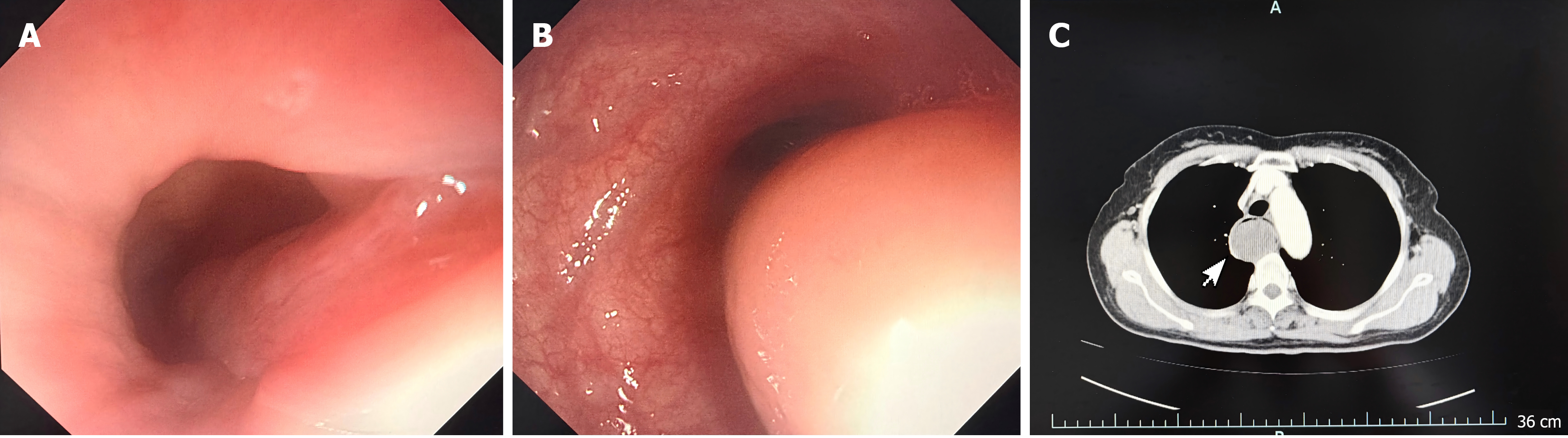
Figure 1 Gastroscopic examination and contrast-enhanced computed tomography.
A: The base of the pedicle: Gastroscopic examination revealed that the tumor pedicle was located at the esophageal inlet, with the tumor prolapsed into the esophagus precluding complete visualization. No obvious active bleeding was observed at the pedicle mucosa, and the surrounding mucosa appeared pale red; B: Main body of the tumor: Gastroscopic examination revealed that the main body of the tumor was round-to-oval in shape, protruding prominently into the esophageal lumen. The surface mucosa of the lesion was intact and smooth, with a pale red color. The vascular pattern of the surrounding esophageal mucosa was clearly visible, and the lumen showed a certain degree of narrowing; C: Contrast-enhanced computed tomography scan of the mediastinum demonstrated a tumor occupying the entire esophageal lumen, with marked esophageal dilatation. The lesion showed homogeneous soft-tissue density, well-defined margins, and no significant enhancement.
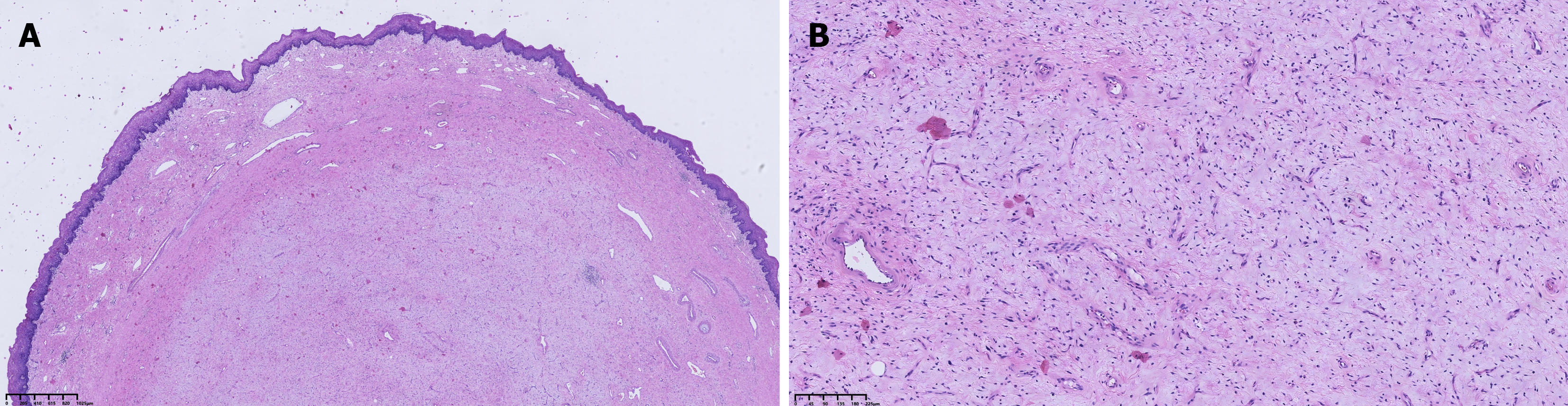
Figure 2 Histopathological examination.
A: Histopathological examination (HE) staining revealed that the tumor surface was covered by stratified squamous epithelium, with marked thickening of the submucosa, which was composed of loose connective tissue, numerous thick-walled vessels, and fibrous tissue, accompanied by prominent stromal edema and myxoid changes (HE staining, objective ×4, scale bar = 1025 μm); B: HE staining revealed submucosal fibrovascular proliferation, with no hyperchromatic atypical lipoblasts or atypical stromal cells identified (HE staining, objective ×10, scale bar = 225 μm).
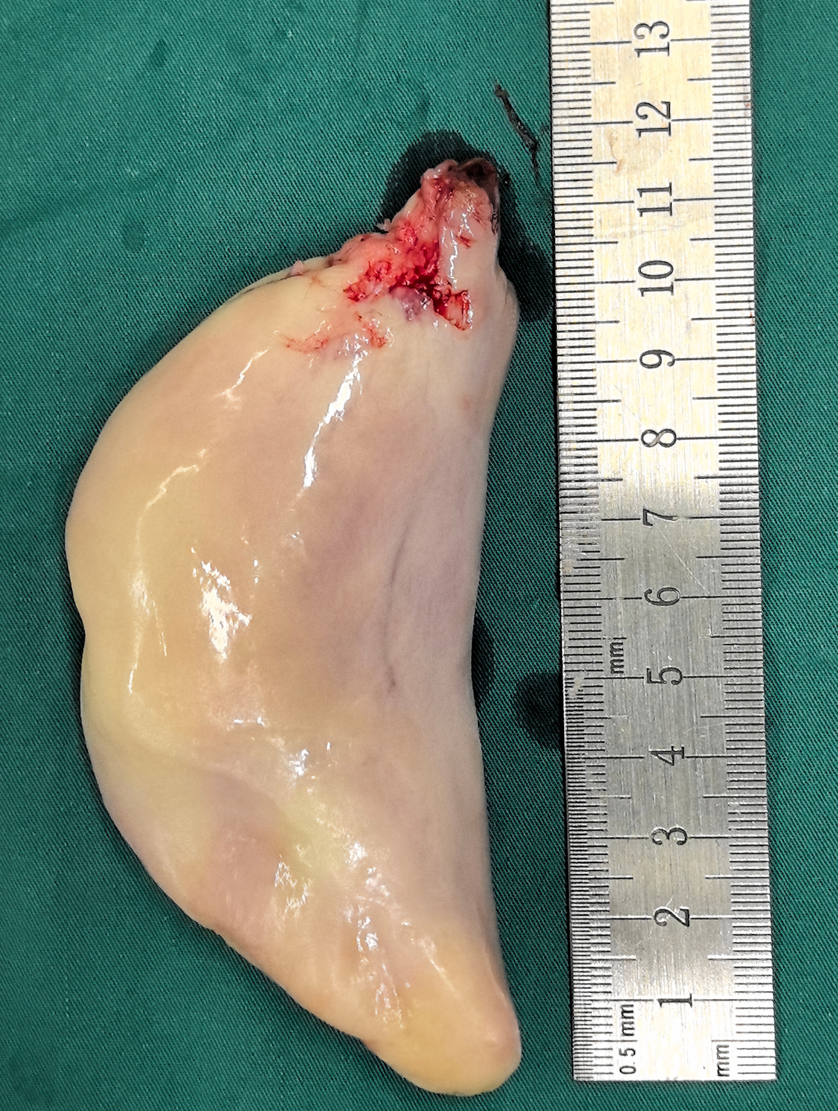
Figure 3 Macroscopic appearance of the tumor: The resected gross specimen was crescent-shaped, with a cut surface showing a white-and-yellow mottled appearance.
At one end, a surgical resection margin with mild hemorrhage was noted. The scale indicated that the maximum diameter of the specimen was approximately 12 cm.
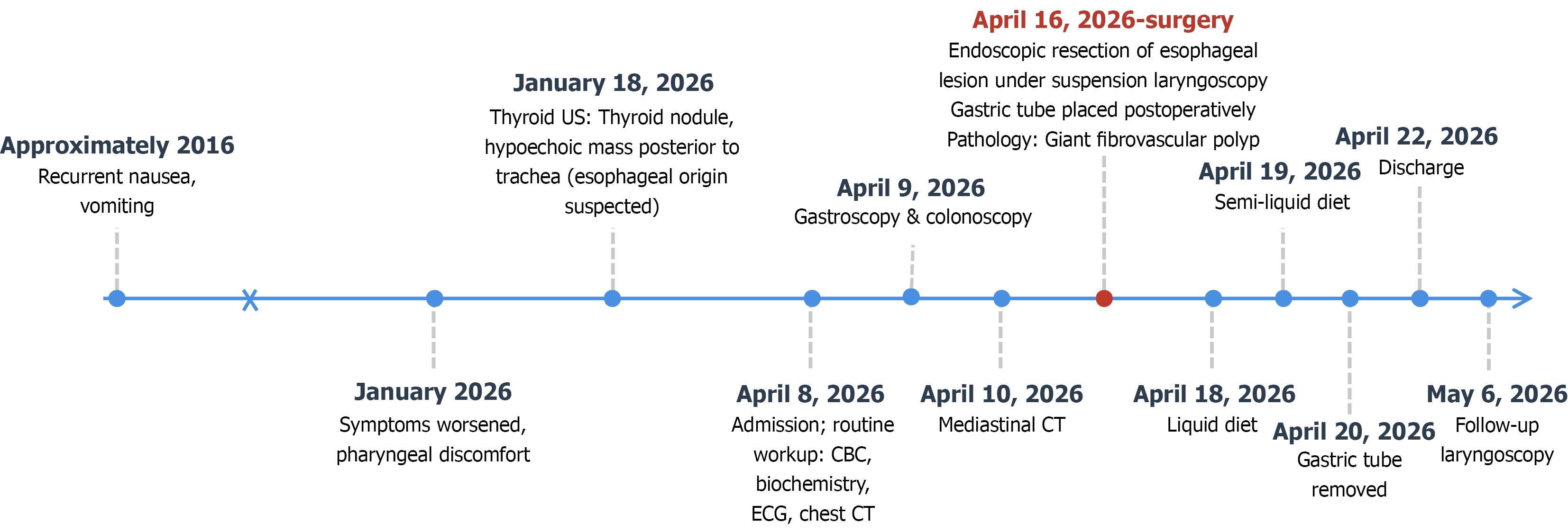
Figure 4 Clinical timeline of the patient: The timeline illustrates the clinical course from symptom onset in approximately 2016 to the follow-up laryngoscopy in May 2026.
Blue circles indicate non-surgical clinical events; the red circle indicates the surgical intervention (endoscopic resection of the esophageal lesion under suspension laryngoscopy). The break in the time axis denotes a large interval between approximately 2016 and January 2026 and is not drawn to scale. CT: Computed tomography; CBC: Complete blood count; ECG: Electrocardiogram; US: Ultrasonography.
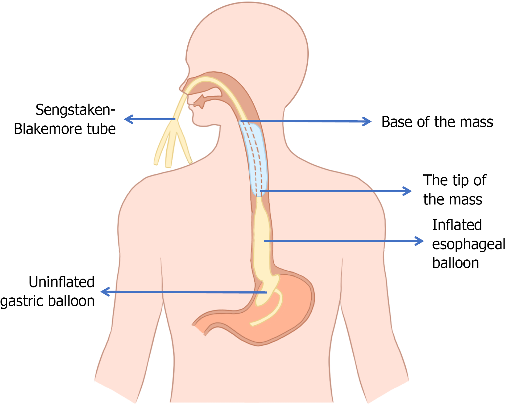
Figure 5 Schematic diagram of the Sengstaken-Blakemore tube placement technique.
The tube is inserted through the nasal cavity, advanced below the mass, and the esophageal balloon is inflated to occlude the esophagus. The tube is then gradually retracted until the balloon contacts the distal end of the mass.
- Citation: Cao MK, Chen XY, Shen QY, Fan Z. Giant esophageal fibrovascular polyp with use of Sengstaken-Blakemore tube during surgery: A case report. World J Gastrointest Endosc 2026; 18(10): 126399
- URL: https://www.wjgnet.com/1948-5190/full/v18/i10/126399.htm
- DOI: https://dx.doi.org/10.4253/wjge.126399