
















Copyright: ©Author(s) 2026.
World J Gastrointest Endosc. Oct 16, 2026; 18(10): 126399
Published online Oct 16, 2026. doi: 10.4253/wjge.126399
Published online Oct 16, 2026. doi: 10.4253/wjge.126399
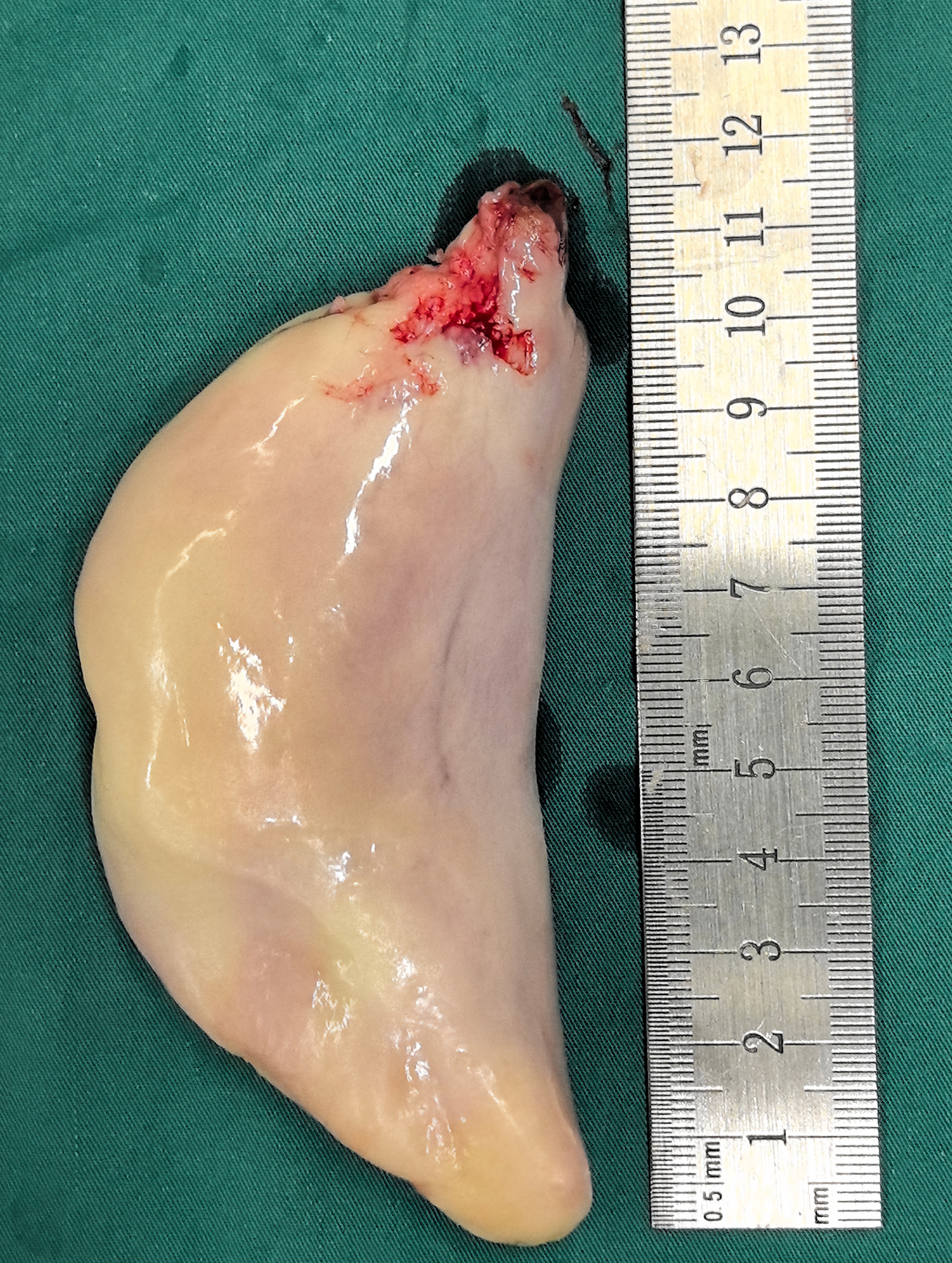
Figure 3 Macroscopic appearance of the tumor: The resected gross specimen was crescent-shaped, with a cut surface showing a white-and-yellow mottled appearance.
At one end, a surgical resection margin with mild hemorrhage was noted. The scale indicated that the maximum diameter of the specimen was approximately 12 cm.
- Citation: Cao MK, Chen XY, Shen QY, Fan Z. Giant esophageal fibrovascular polyp with use of Sengstaken-Blakemore tube during surgery: A case report. World J Gastrointest Endosc 2026; 18(10): 126399
- URL: https://www.wjgnet.com/1948-5190/full/v18/i10/126399.htm
- DOI: https://dx.doi.org/10.4253/wjge.126399