
















Copyright: ©Author(s) 2026.
World J Gastrointest Endosc. Oct 16, 2026; 18(10): 126399
Published online Oct 16, 2026. doi: 10.4253/wjge.126399
Published online Oct 16, 2026. doi: 10.4253/wjge.126399
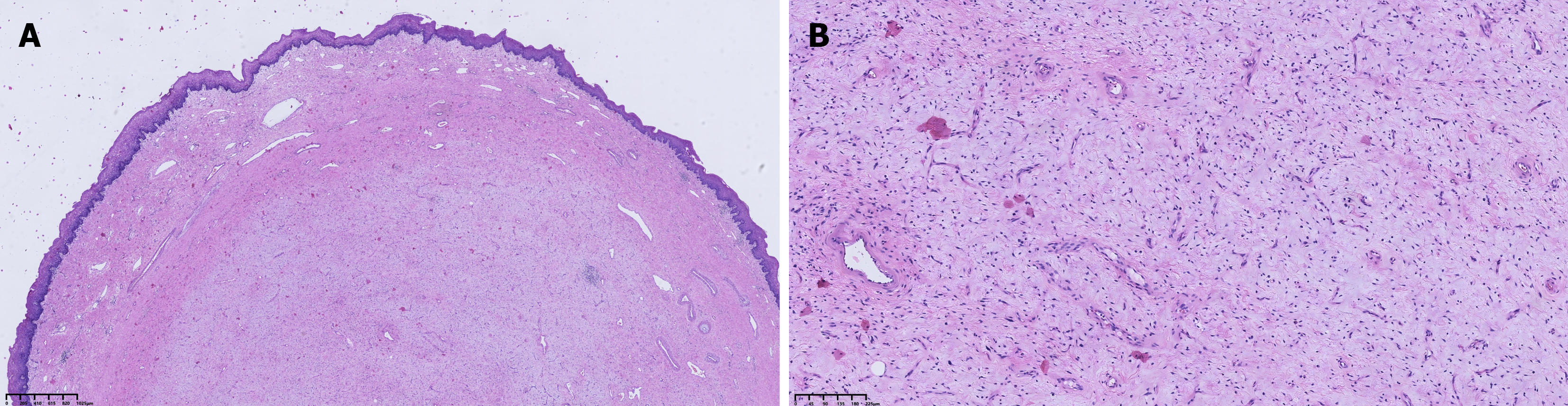
Figure 2 Histopathological examination.
A: Histopathological examination (HE) staining revealed that the tumor surface was covered by stratified squamous epithelium, with marked thickening of the submucosa, which was composed of loose connective tissue, numerous thick-walled vessels, and fibrous tissue, accompanied by prominent stromal edema and myxoid changes (HE staining, objective ×4, scale bar = 1025 μm); B: HE staining revealed submucosal fibrovascular proliferation, with no hyperchromatic atypical lipoblasts or atypical stromal cells identified (HE staining, objective ×10, scale bar = 225 μm).
- Citation: Cao MK, Chen XY, Shen QY, Fan Z. Giant esophageal fibrovascular polyp with use of Sengstaken-Blakemore tube during surgery: A case report. World J Gastrointest Endosc 2026; 18(10): 126399
- URL: https://www.wjgnet.com/1948-5190/full/v18/i10/126399.htm
- DOI: https://dx.doi.org/10.4253/wjge.126399