
















Copyright: ©Author(s) 2026.
World J Gastrointest Endosc. Oct 16, 2026; 18(10): 126399
Published online Oct 16, 2026. doi: 10.4253/wjge.126399
Published online Oct 16, 2026. doi: 10.4253/wjge.126399
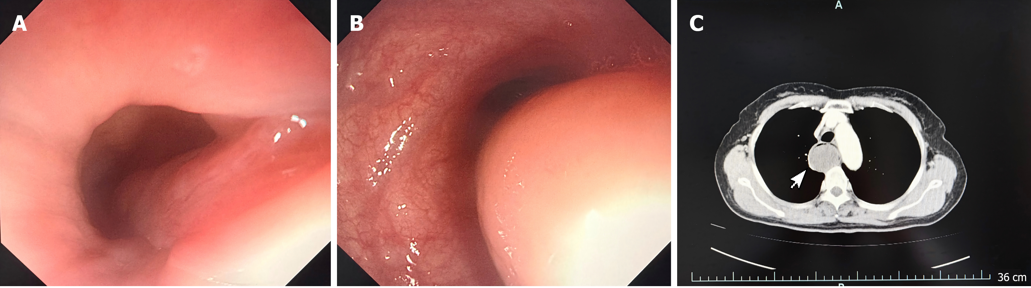
Figure 1 Gastroscopic examination and contrast-enhanced computed tomography.
A: The base of the pedicle: Gastroscopic examination revealed that the tumor pedicle was located at the esophageal inlet, with the tumor prolapsed into the esophagus precluding complete visualization. No obvious active bleeding was observed at the pedicle mucosa, and the surrounding mucosa appeared pale red; B: Main body of the tumor: Gastroscopic examination revealed that the main body of the tumor was round-to-oval in shape, protruding prominently into the esophageal lumen. The surface mucosa of the lesion was intact and smooth, with a pale red color. The vascular pattern of the surrounding esophageal mucosa was clearly visible, and the lumen showed a certain degree of narrowing; C: Contrast-enhanced computed tomography scan of the mediastinum demonstrated a tumor occupying the entire esophageal lumen, with marked esophageal dilatation. The lesion showed homogeneous soft-tissue density, well-defined margins, and no significant enhancement.
- Citation: Cao MK, Chen XY, Shen QY, Fan Z. Giant esophageal fibrovascular polyp with use of Sengstaken-Blakemore tube during surgery: A case report. World J Gastrointest Endosc 2026; 18(10): 126399
- URL: https://www.wjgnet.com/1948-5190/full/v18/i10/126399.htm
- DOI: https://dx.doi.org/10.4253/wjge.126399