
















Copyright: ©Author(s) 2026.
World J Gastrointest Endosc. Oct 16, 2026; 18(10): 126399
Published online Oct 16, 2026. doi: 10.4253/wjge.126399
Published online Oct 16, 2026. doi: 10.4253/wjge.126399
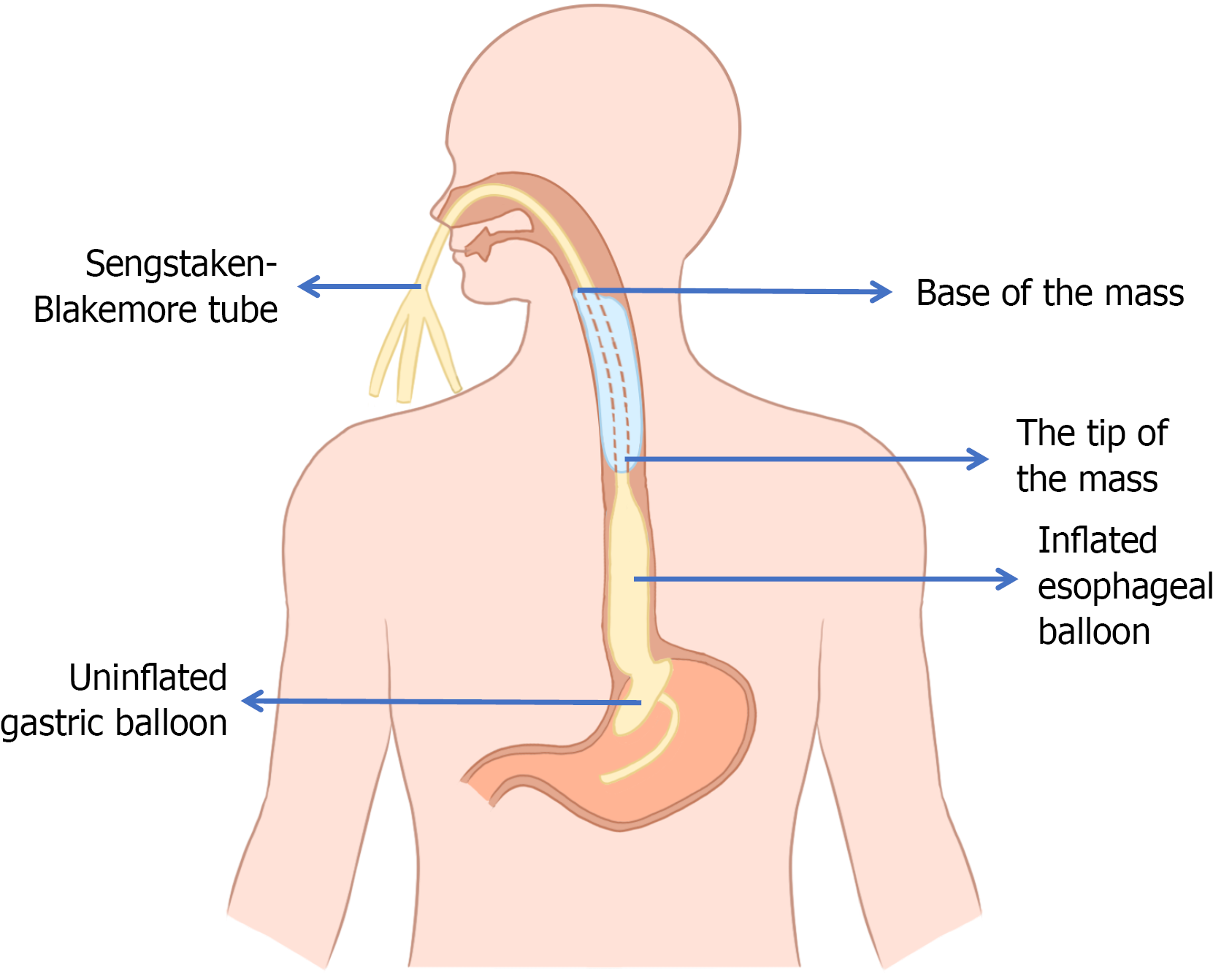
Figure 5 Schematic diagram of the Sengstaken-Blakemore tube placement technique.
The tube is inserted through the nasal cavity, advanced below the mass, and the esophageal balloon is inflated to occlude the esophagus. The tube is then gradually retracted until the balloon contacts the distal end of the mass.
- Citation: Cao MK, Chen XY, Shen QY, Fan Z. Giant esophageal fibrovascular polyp with use of Sengstaken-Blakemore tube during surgery: A case report. World J Gastrointest Endosc 2026; 18(10): 126399
- URL: https://www.wjgnet.com/1948-5190/full/v18/i10/126399.htm
- DOI: https://dx.doi.org/10.4253/wjge.126399