
















Copyright: ©Author(s) 2026.
World J Stem Cells. Sep 26, 2026; 18(9): 121744
Published online Sep 26, 2026. doi: 10.4252/wjsc.121744
Published online Sep 26, 2026. doi: 10.4252/wjsc.121744
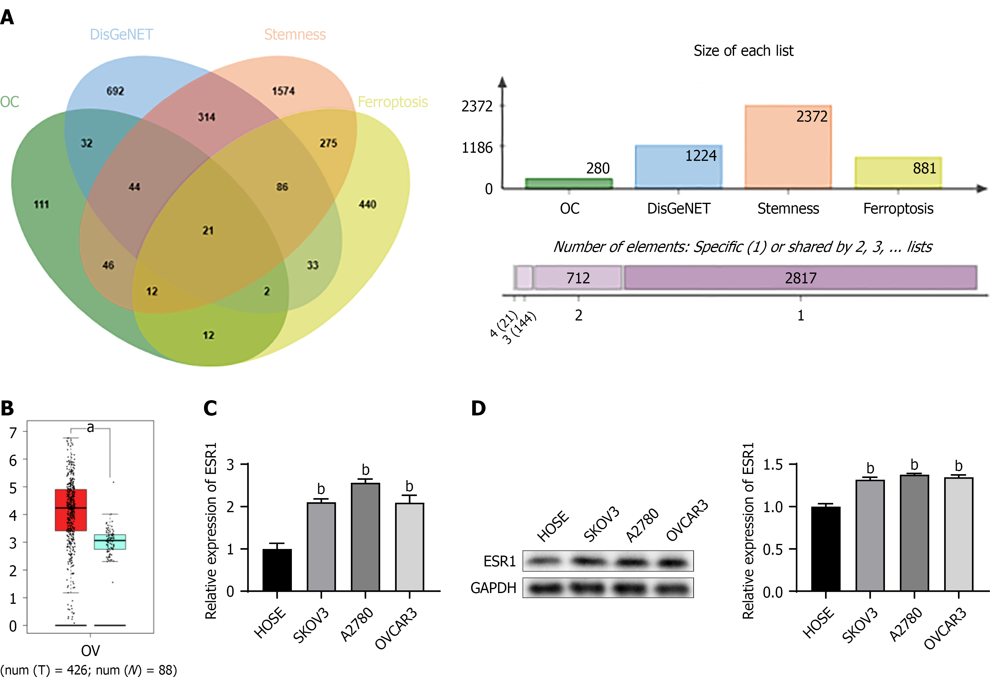
Figure 1 Expression of estrogen receptor 1 in ovarian cancer patients and cell lines.
A: Screening of targeted genes associated with ovarian cancer, cell stemness, and ferroptosis using DisGeNET and GeneCards databases; B: GEPIA analysis of estrogen receptor 1 (ESR1) in ovarian cancer patients and health controls; C: Real-time polymerase chain reaction analysis revealed increased ESR1 levels in ovarian cancer cell lines; D: Western blot analysis revealed increased ESR1 levels. Data was expressed as mean ± SD (n = 3). aP < 0.01; bP < 0.01 vs HOSE group. ESR1: Estrogen receptor 1.
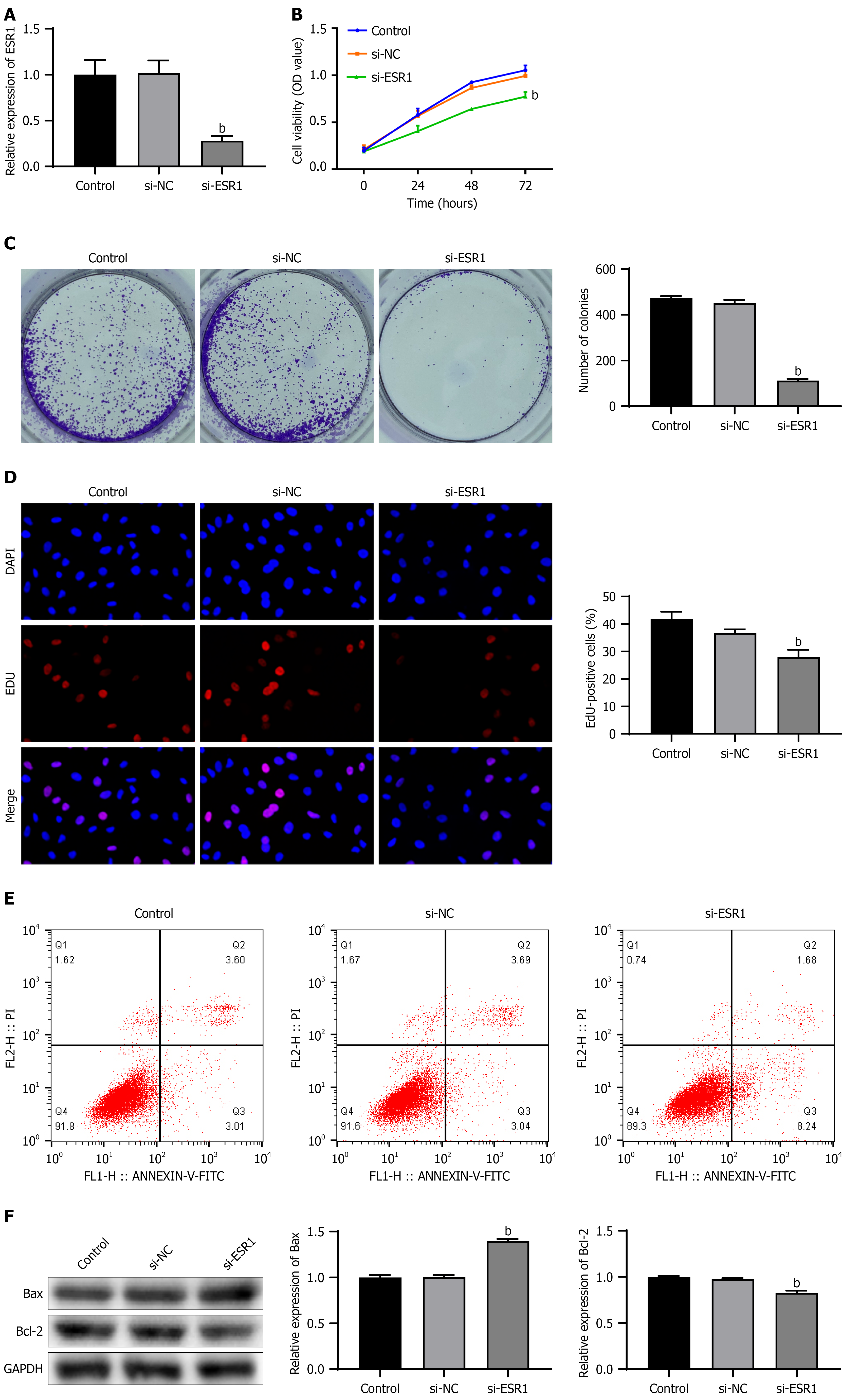
Figure 2 Effect of estrogen receptor 1 downregulation on cell function in A2780 cells.
A: Real-time polymerase chain reaction analysis revealed a significant decrease in estrogen receptor 1 (ESR1) levels in A2780 cells following transfection with si-ESR1. Then, A2780 cells were transfected with si-NC and si-ESR1; B: Cell viability was notably reduced by Cell Counting Kit-8 assay; C: Cell proliferation was notably reduced by colony formation assay; D: Cell proliferation was notably reduced by 5-ethynyl-2’-deoxyuridine assay; E: Cell apoptosis was increased by flow cytometry; F: Expression levels of B-cell lymphoma-2 associated X were significantly elevated, while B-cell lymphoma-2 expression was decreased. Data was expressed as mean ± SD (n = 3). bP < 0.01 vs si-NC group. ESR1: Estrogen receptor 1; EDU: 5-ethynyl-2’-deoxyuridine assay; Bcl-2: B-cell lymphoma-2; Bax: B-cell lymphoma-2 associated X.
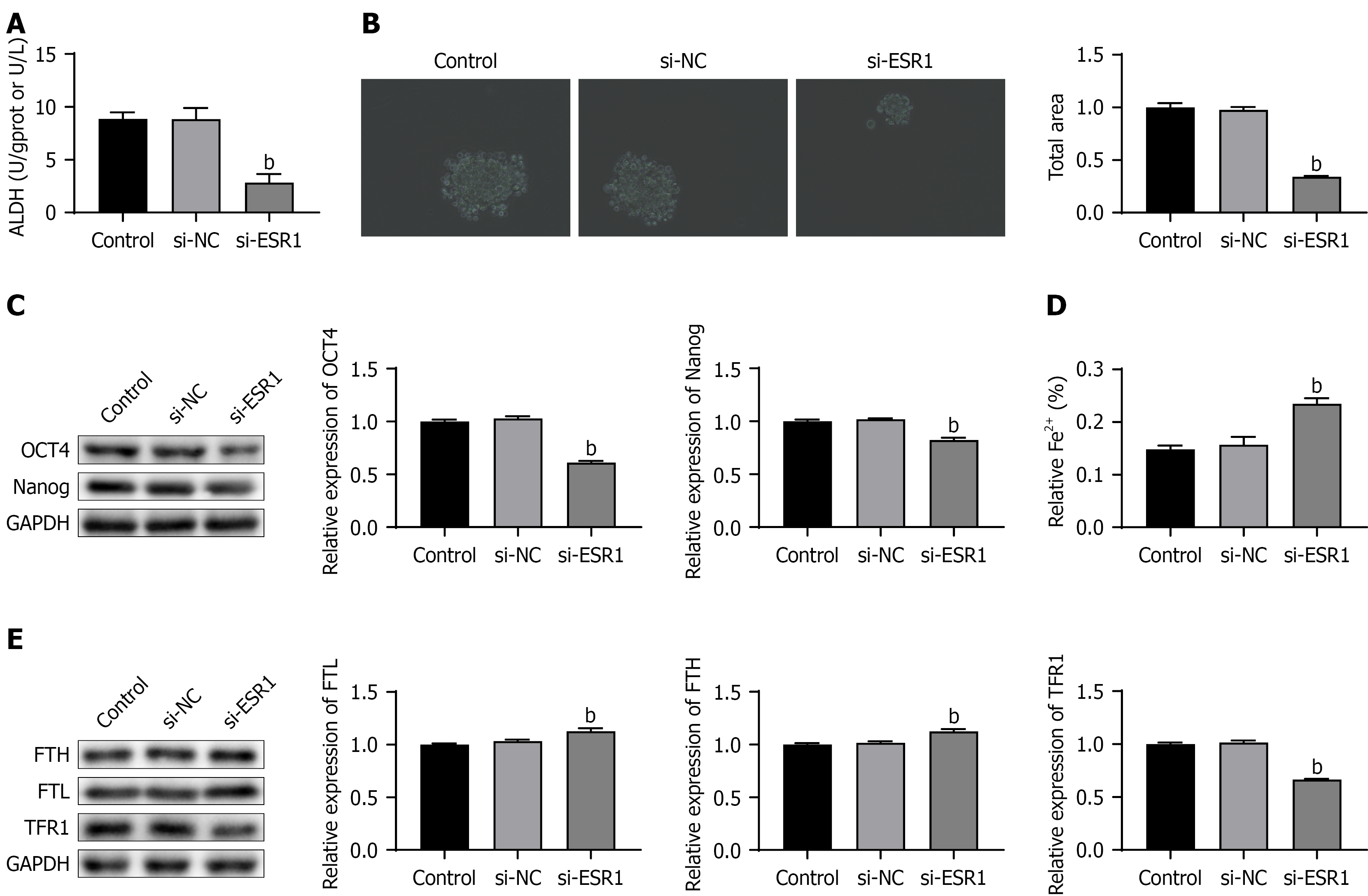
Figure 3 Effect of estrogen receptor 1 downregulation on cell stemness and ferroptosis in A2780 cells.
A2780 cells were transfected with si-NC and si-ESR1. A: Aldehyde dehydrogenas activity was reduced following transfection with si-ESR1; B: Cell spheroidization assay demonstrated a decrease in cell stemness; C: Protein levels of octamer-binding transcription factor 4 and Nanog were notably decreased; D: Fe2+ content increased; E: Expression levels of ferritin heavy chain and ferritin light chain were significantly elevated, while the expression of transferrin receptor 1 was decreased. Data was expressed as mean ± SD (n = 3). bP < 0.01 vs si-NC group. ALDH: Aldehyde dehydrogenas; ESR1: Estrogen receptor 1; OCT4: Octamer-binding transcription factor 4; FTH: Ferritin heavy chain; FTL: Ferritin light chain; TFR1: Transferrin receptor 1.
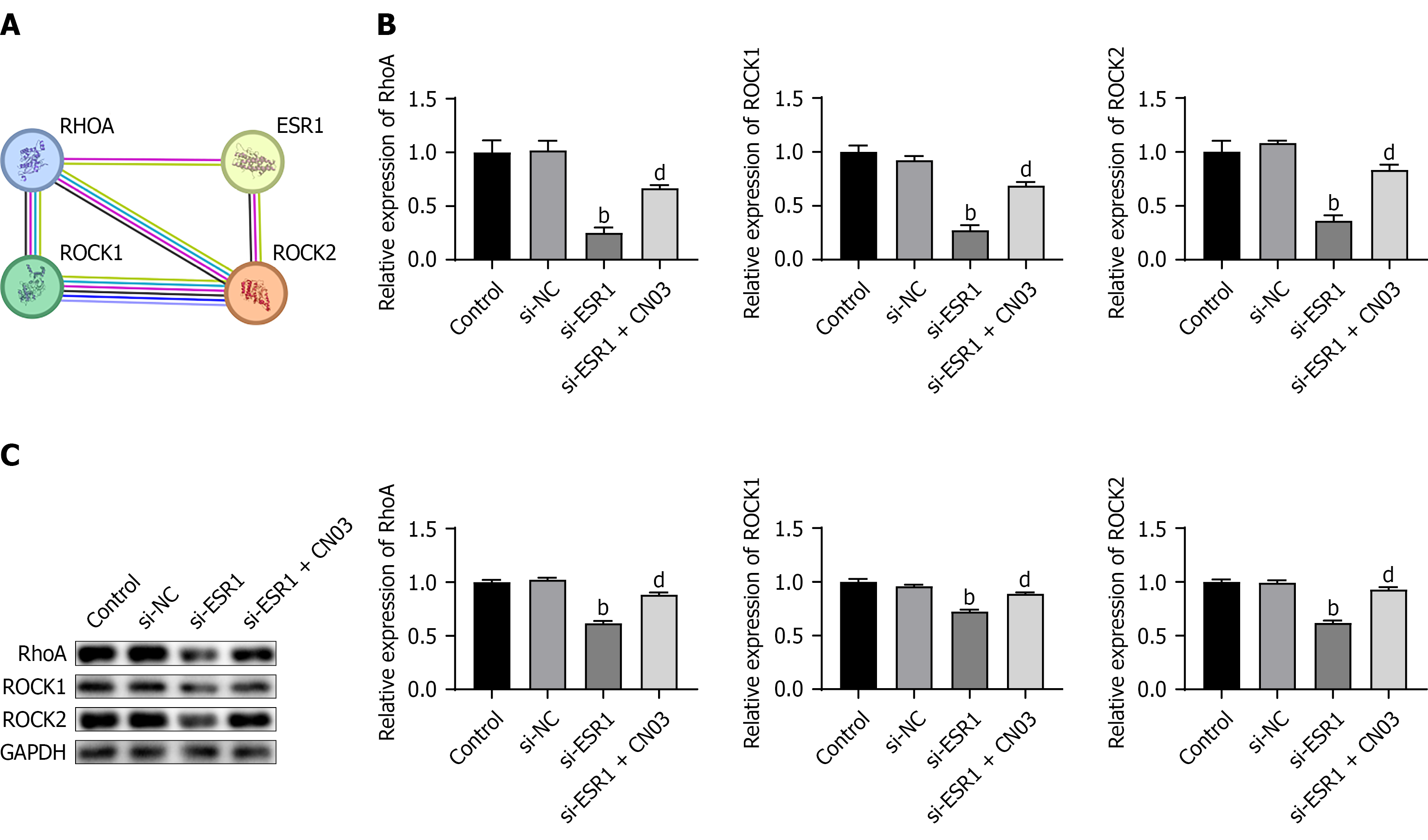
Figure 4 Effect of estrogen receptor 1 downregulation on the Ras homolog gene family member A/Rho-associated coiled-coil containing protein kinase signaling pathway in A2780 cells.
A: Protein-protein interaction network analysis. A2780 cells were transfected with si-NC and si-ESR1, and treated with CN03; B: Real-time polymerase chain reaction analyzed the mRNA levels of RhoA, ROCK1, and ROCK2 in A2780 cells transfected with si-ESR1 with or without CN03; C: Western blot analyzed the protein levels of RhoA, ROCK1, and ROCK2. Data was expressed as mean ± SD (n = 3). bP < 0.01 vs si-NC group; dP < 0.01 vs si-ESR1 group. ESR1: Estrogen receptor 1; RHOA: Ras homolog gene family member A; ROCK: Rho-associated coiled-coil containing protein kinase.
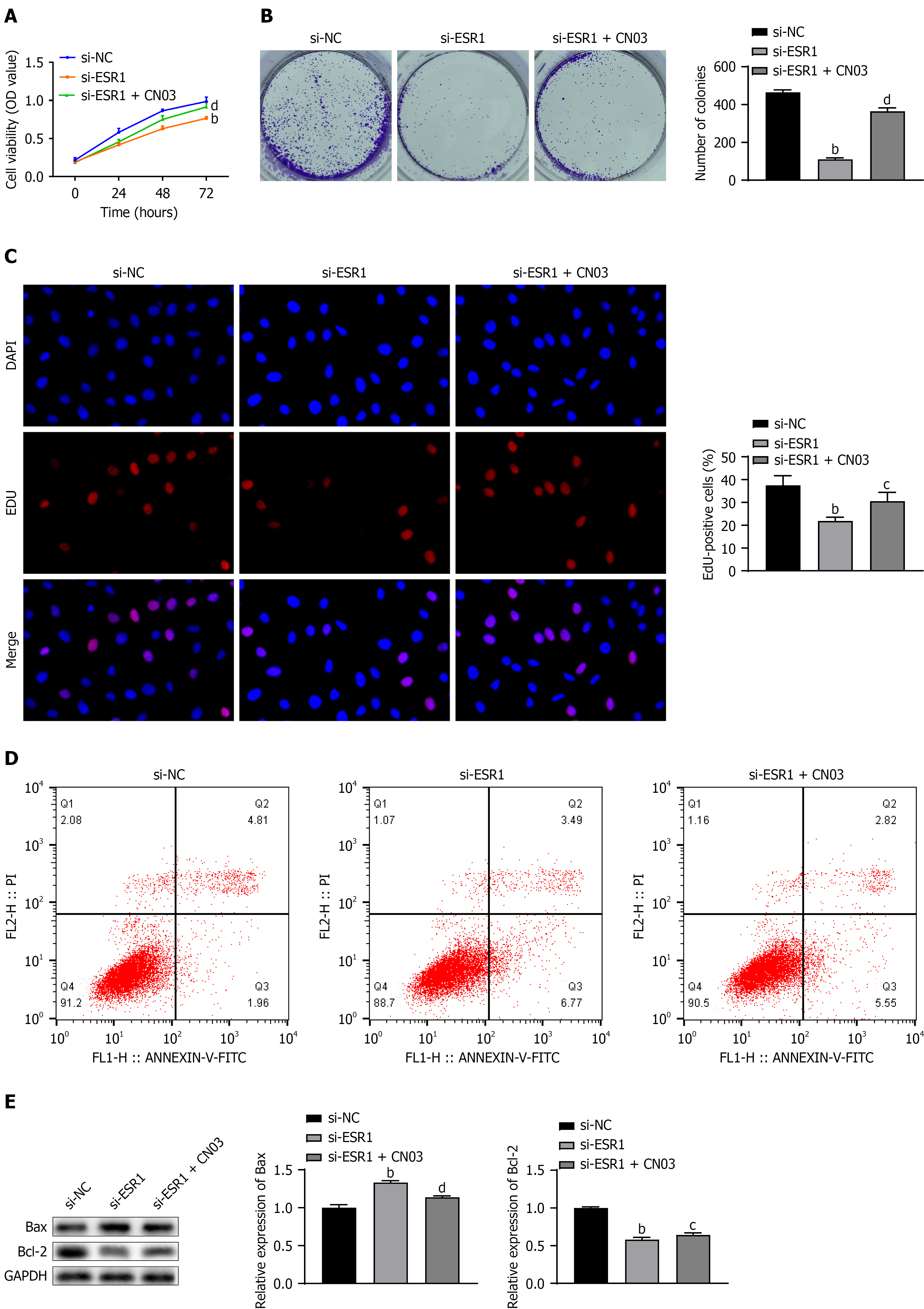
Figure 5 Effect of estrogen receptor 1 downregulation on cell function in A2780 cells by inhibiting the Ras homolog gene family member A/Rho-associated coiled-coil containing protein kinase signaling pathway.
A2780 cells were transfected with si-NC and si-ESR1, and treated with CN03. A: Cell viability significantly decreased in A2780 cells following transfection with si-ESR1, which was rescued by CN03 treatment; B: Colony formation assay showed reduced cell proliferation in the si-ESR1 group, with restoration upon CN03 introduction; C: 5-ethynyl-2’-deoxyuridine assay revealed decreased cell proliferation in the si-ESR1 group, which was alleviated by CN03 treatment; D: Flow cytometry analysis demonstrated increased apoptosis in A2780 cells post si-ESR1 transfection, with apoptosis inhibition observed in the si-ESR1+ CN03 group; E: Expression levels of B-cell lymphoma-2 associated X and B-cell lymphoma-2 were altered in the si-ESR1 group, with an increase in B-cell lymphoma-2 associated X expression and a decrease in B-cell lymphoma-2 expression, both of which were reversed by CN03 treatment. Data was expressed as mean ± SD (n = 3). bP < 0.01 vs si-NC group; cP < 0.05 vs si-ESR1 group; dP < 0.01 vs si-ESR1 group. ESR1: Estrogen receptor 1; EDU: 5-ethynyl-2’-deoxyuridine assay; Bcl-2: B-cell lymphoma-2; Bax: B-cell lymphoma-2 associated X.
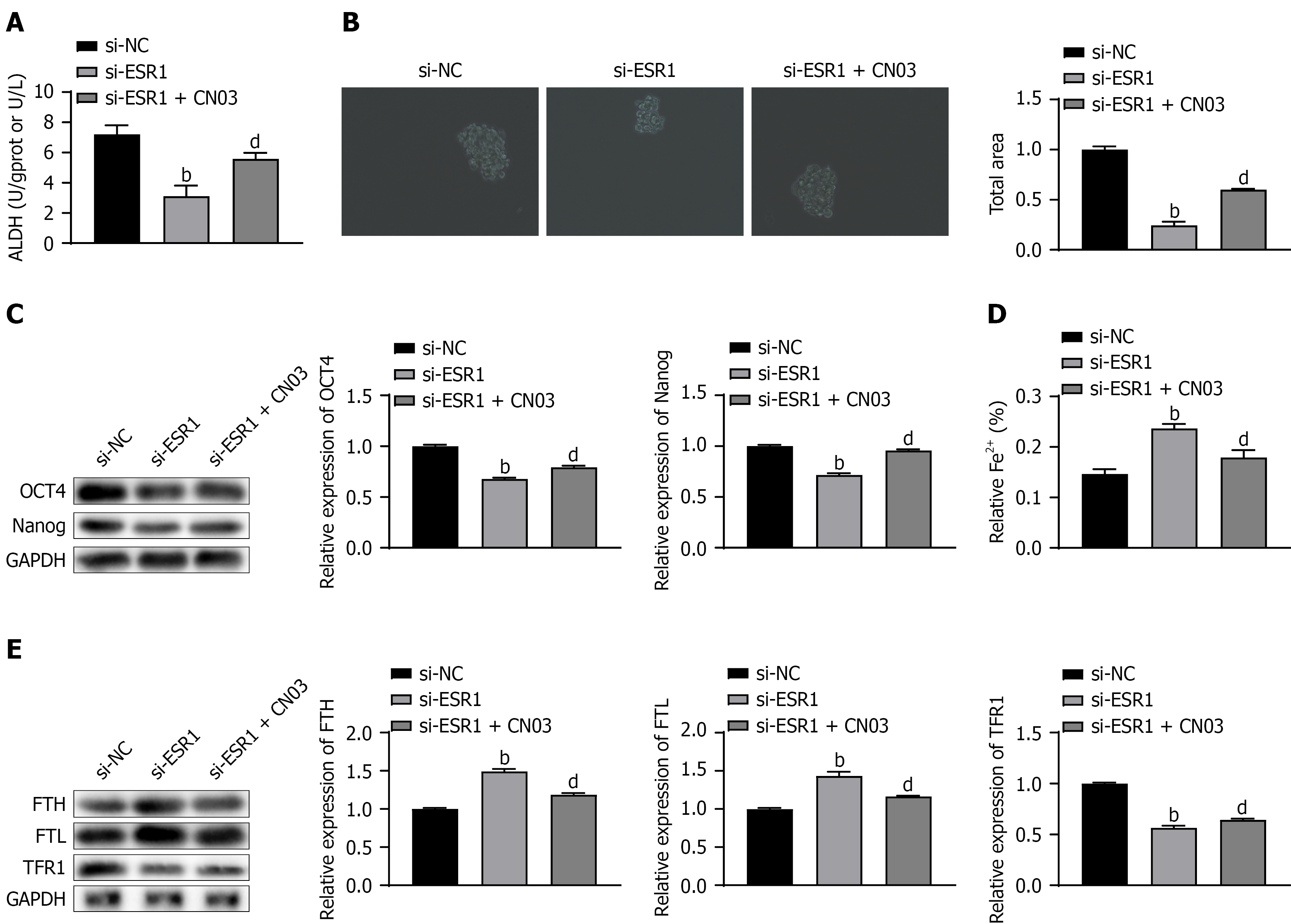
Figure 6 Effect of estrogen receptor 1 downregulation on cell stemness and ferroptosis in A2780 cells by inhibiting the Ras homolog gene family member A/Rho-associated coiled-coil containing protein kinase signaling pathway.
A2780 cells were transfected with si-NC and si-ESR1, and treated with CN03. A: Aldehyde dehydrogenas activity significantly decreased in A2780 cells post si-ESR1 transfection, which was partially restored by CN03 treatment; B: Cell spheroidization assay demonstrated a decrease in cell stemness in the si-ESR1 group, with a reversal observed upon CN03 introduction; C: Protein levels of octamer-binding transcription factor 4 and Nanog decreased significantly in the si-ESR1 group, but were restored by CN03 treatment; D: Fe2+ content in A2780 cells increased following si-ESR1 transfection, and decreased with CN03 supplementation; E: Expression levels of ferritin heavy chain and ferritin light chain increased, while transferrin receptor 1 expression decreased in the si-ESR1 group, and these changes were reversed by CN03 treatment. Data was expressed as mean ± SD (n = 3). bP < 0.01 vs si-NC group; dP < 0.01 vs si-ESR1 group. ALDH: Aldehyde dehydrogenas; ESR1: Estrogen receptor 1; OCT4: Octamer-binding transcription factor 4; FTH: Ferritin heavy chain; FTL: Ferritin light chain; TFR1: Transferrin receptor 1.
- Citation: Zhou S, Cheng X, Zhang JM, Ji J, Ni ZZ, Wang W. Estrogen receptor 1 regulates ovarian cancer cell stemness and ferroptosis through modulation of the canonical RhoA/ROCK signaling pathway. World J Stem Cells 2026; 18(9): 121744
- URL: https://www.wjgnet.com/1948-0210/full/v18/i9/121744.htm
- DOI: https://dx.doi.org/10.4252/wjsc.121744