Copyright
©The Author(s) 2018.
World J Gastroenterol. Mar 7, 2018; 24(9): 992-1003
Published online Mar 7, 2018. doi: 10.3748/wjg.v24.i9.992
Published online Mar 7, 2018. doi: 10.3748/wjg.v24.i9.992
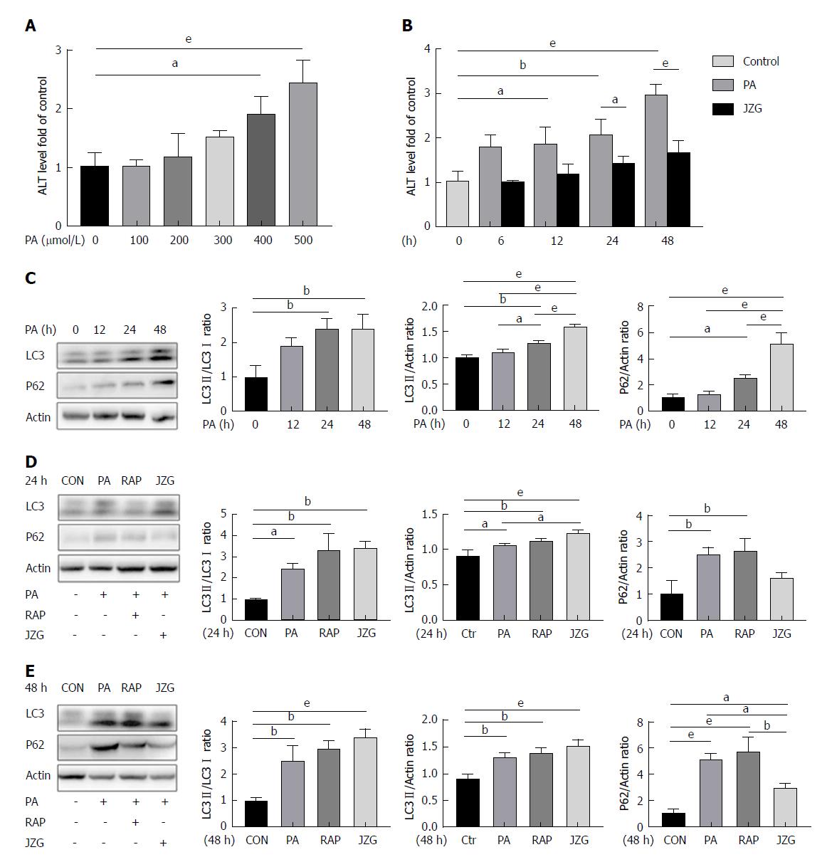
Figure 1 Autophagy was activated by JZG to protect against injury in palmitate-treated cells.
A: The expression of ALT in the supernatants of cell cultures was determined by ELISA. HepG2 cells were incubated in culture medium containing PA (0, 0.1, 0.2, 0.3, 0.4 or 0.5 mmol/L); B: The expression of ALT in the supernatants of cell cultures was determined by ELISA. HepG2 cells were treated with 0.4 mmol/L PA with or without JZG (100 μg/mL) for different time periods (0, 6, 12, 24 or 48 h); C: HepG2 cells were treated with 0.4 mmol/L PA for different time periods (0, 12, 24 or 48 h); D: HepG2 cells were treated with 0.4 mmol/L PA and rapamycin (2 μmol/L) or JZG (100 μg/mL) for 24 h; E: HepG2 cells were treated with 0.4 mmol/L PA and rapamycin (2 μmol/L) or JZG (100 μg/mL) for 48 h. Data are expressed as mean ± SEM. aP < 0.05, bP < 0.01, eP < 0.001. ALT: Alanine aminotransferase; ELISA: Enzyme-linked immunosorbent assay; JZG: Jiang Zhi Granule; PA: Palmitate.
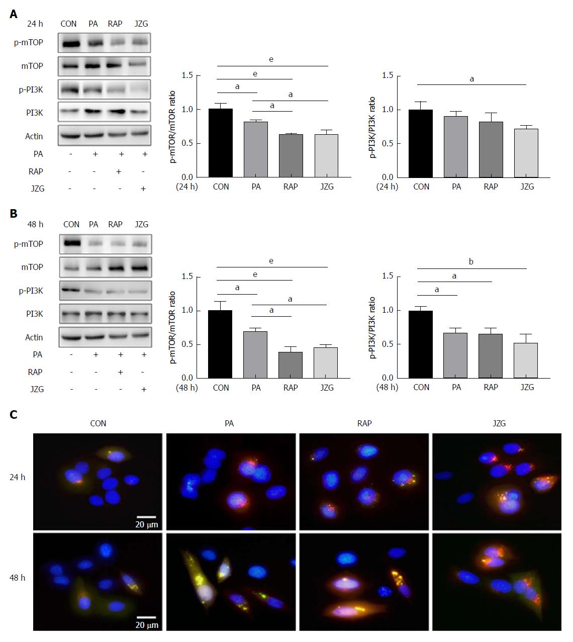
Figure 2 PI3K-AKT-mTOR pathway was involved in autophagy in Jiang Zhi Granule-treated cells.
A: HepG2 cells were treated with 0.4 mmol/L PA and rapamycin (2 μmol/L) or JZG (100 μg/mL) for 24 h; B: HepG2 cells were treated with 0.4 mmol/L PA and rapamycin (2 μmol/L) or JZG (100 μg/mL) for 48 h; C: HepG2 cells stably expressing mRFP-GFP-LC3 were pretreated with 0.4 mmol/L PA and rapamycin (2 μmol/L) or JZG (100 μg/mL) for 24 h and 48 h, and then analyzed by fluorescence microscopy. Data are expressed as mean ± SEM. aP < 0.05, bP < 0.01, eP < 0.001. JZG: Jiang Zhi Granule; PA: Palmitate.
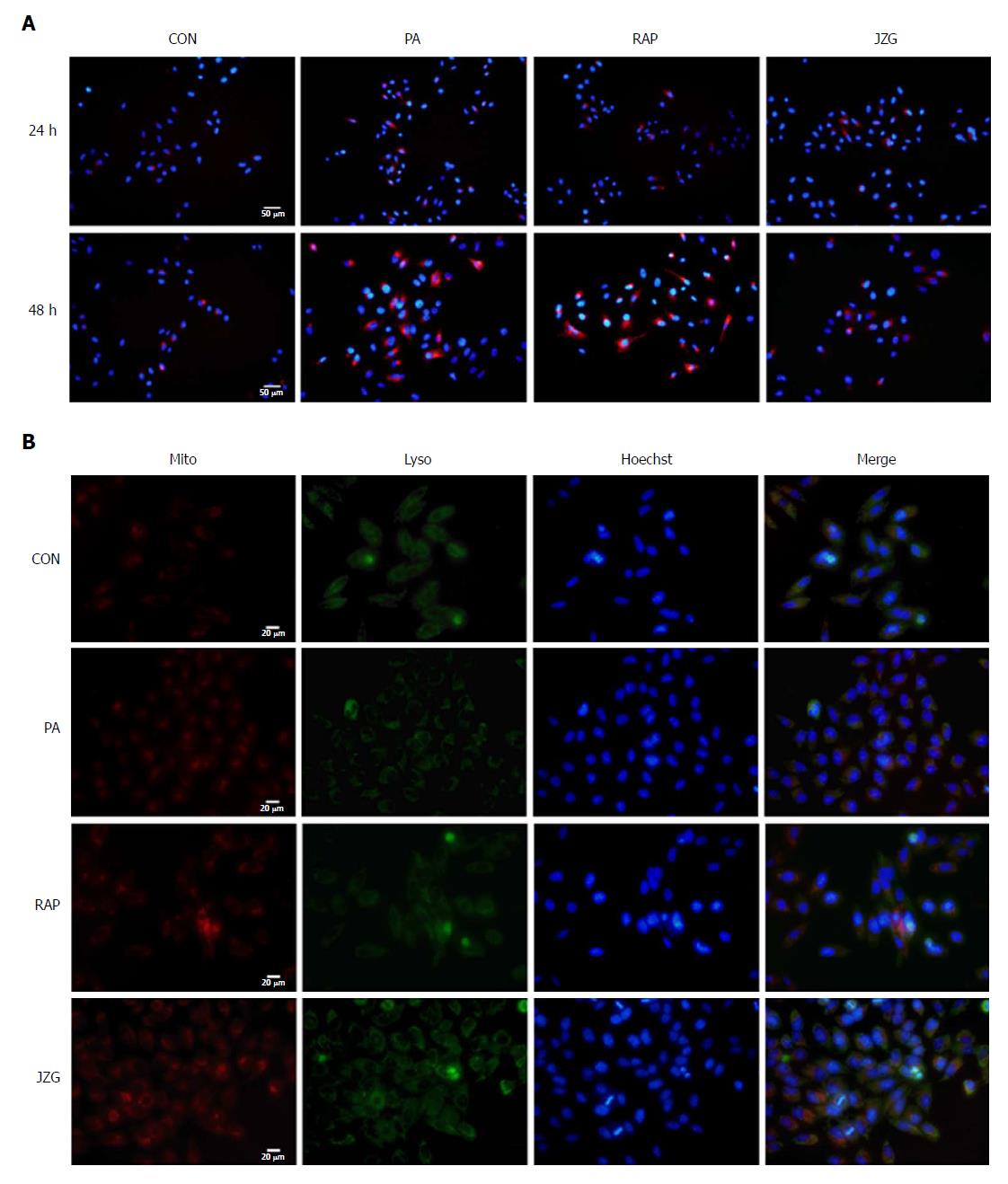
Figure 3 Effects of Jiang Zhi Granule on autophagic flux in palmitate-treated cells.
A: HepG2 cells stably expressing mCherry-p62 were pretreated with 0.4 mmol/L PA and rapamycin (2 μmol/L) or JZG (100 μg/mL) for 24 h and 48 h, and then analyzed by fluorescence microscopy; B: HepG2 cells were treated with 0.4 mmol/L PA and rapamycin (2 μmol/L) or JZG (100 μg/mL) for 48 h, and then analyzed by fluorescence microscopy. JZG: Jiang Zhi Granule; PA: Palmitate.
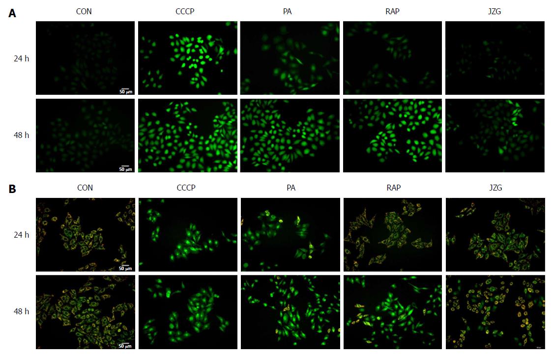
Figure 4 Jiang Zhi Granule protected mitochondrial integrity against oxidative stress.
A: The ROS-sensitive fluorescent probe DCFH-DA was used to monitor mitochondrial membrane potential in HepG2 cells pretreated with 0.4 mmol/L PA and rapamycin (2 μmol/L) or JZG (100 μg/mL) for 24 h and 48 h, and the cells were then analyzed by fluorescence microscopy; B: JC-1 was used to monitor mitochondrial membrane potential in HepG2 cells pretreated with 0.4 mmol/L PA and rapamycin (2 μmol/L) or JZG (100 μg/mL) for 24 h and 48 h, and the cells were then analyzed by fluorescence microscopy. JZG: Jiang Zhi Granule; PA: Palmitate; ROS: Reactive oxygen species.
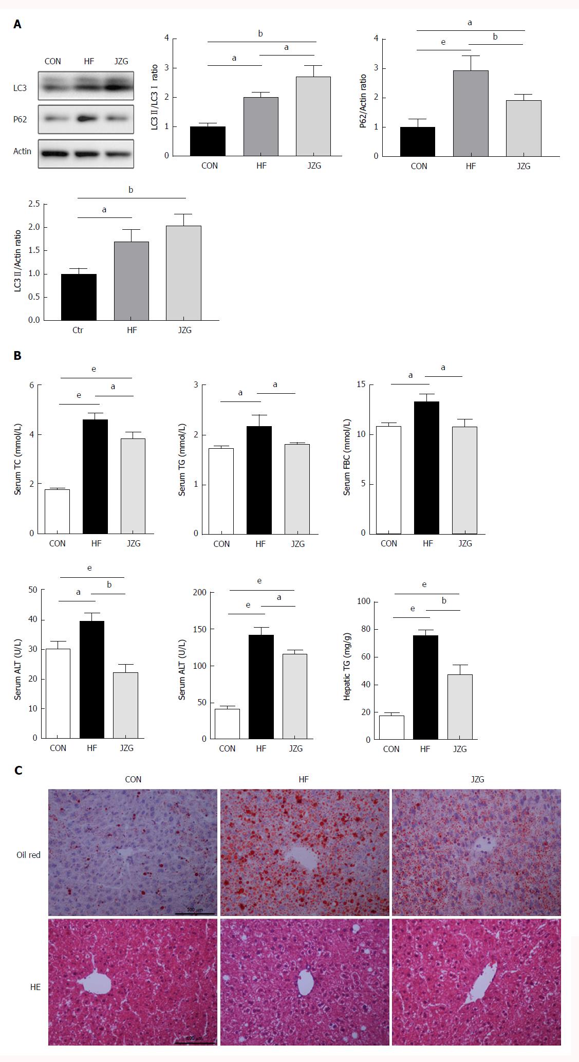
Figure 5 Autophagy was activated by Jiang Zhi Granule to improve non-alcoholic fatty liver disease in vivo.
A: Autophagy of liver tissue was examined by western blot analyses; B: Biochemical analysis of TG, TC, ALT, AST and FBG using an automatic blood chemistry analyzer; C: Inflammation and lipid content in the liver was detected by HE staining and oil Red O staining. Data are expressed as mean ± SEM. aP < 0.05, bP < 0.01, eP < 0.001. ALT: Alanine aminotransferase; AST: Aspartate aminotransferase; HE: Hematoxylin and eosin; JZG: Jiang Zhi Granule; PA: Palmitate; TC: Total cholesterol; TG: Triglycerides.
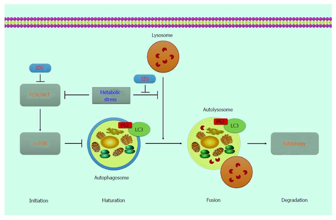
Figure 6 The potential role of autophagy in metabolic stress-induced hepatocyte injury and the protective mechanism of JZG in this process.
JZG: Jiang Zhi Granule.
- Citation: Zheng YY, Wang M, Shu XB, Zheng PY, Ji G. Autophagy activation by Jiang Zhi Granule protects against metabolic stress-induced hepatocyte injury. World J Gastroenterol 2018; 24(9): 992-1003
- URL: https://www.wjgnet.com/1007-9327/full/v24/i9/992.htm
- DOI: https://dx.doi.org/10.3748/wjg.v24.i9.992


