Copyright
©The Author(s) 2017.
World J Gastroenterol. Sep 21, 2017; 23(35): 6540-6545
Published online Sep 21, 2017. doi: 10.3748/wjg.v23.i35.6540
Published online Sep 21, 2017. doi: 10.3748/wjg.v23.i35.6540
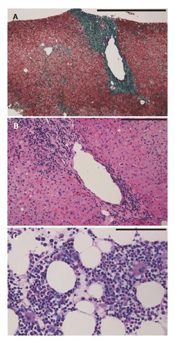
Figure 1 Histopathological features of liver biopsy specimen and clot section of bone marrow aspirate.
A: The liver biopsy specimen shows fibrous portal expansion. There is no fibrous bridging (Elastica-Goldner staining, scale bar; 500 μm); B: Mild piecemeal necrosis, mild intralobular degeneration and focal necrosis, and moderate portal inflammation are observed (H and E staining, scale bar; 200 μm); C: The clot section of bone marrow aspirate shows normal numbers of megakaryocytes and other cell lineages are preserved (Periodic Acid Schiff staining, scale bar; 100 μm).
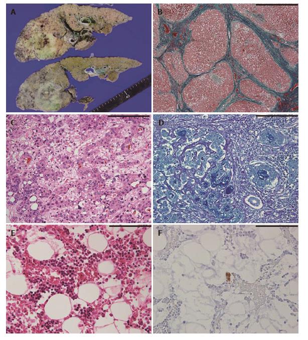
Figure 2 Macroscopic and histopathological features of autopsy specimens.
A: The cut surface of the liver shows diffuse micronodular cirrhosis with a yellow-green lesion in the right lobe; B: The non-tumorous liver shows diffuse small regenerative nodules with fibrous septum (Elastica-Goldner staining, scale bar; 1000 μm); C and D: Histopathological findings of combined hepatocellular-cholangiocarcinoma; C: hepatocellular carcinoma component (H and E staining) and D: adenocarcinoma component (Alcian Blue-Periodic Acid Schiff staining) (C and D, scale bar; 200 μm); E: In the bone marrow, no megakaryocytes are observed (H and E staining); F: A small megakaryocyte is identified through immunostaining for CD41 (E and F, scale bar; 100 μm).
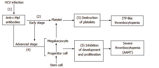
Figure 3 A schema of the pathogenesis of the current case.
Hepatitis C virus infection may cause the generation of anti-c-Mpl antibodies (1). At first, most of the generated antibodies would be absorbed with the c-Mpl on platelets because platelets are the largest component in the megakaryocyte lineage (2). These antibody-attached platelets are destroyed in the spleen (3), therefore, idiopathic thrombocytopenic purpura (ITP)-like clinical manifestations are observed (Early stage). Following a sufficient reduction of platelets, these antibodies begin to bind to the c-Mpl on the megakaryocytes and its progenitor cells in the bone marrow (4). Attached antibodies block the functions of thrombopoietin, causing inhibition in the development and proliferation of the megakaryocyte lineage (5). Thus, severe reduction of megakaryocytes in the bone marrow occurs, that is, acquired amegakaryocytic thrombocytopenia (AAMT) (Advanced stage).
- Citation: Ichimata S, Kobayashi M, Honda K, Shibata S, Matsumoto A, Kanno H. Acquired amegakaryocytic thrombocytopenia previously diagnosed as idiopathic thrombocytopenic purpura in a patient with hepatitis C virus infection. World J Gastroenterol 2017; 23(35): 6540-6545
- URL: https://www.wjgnet.com/1007-9327/full/v23/i35/6540.htm
- DOI: https://dx.doi.org/10.3748/wjg.v23.i35.6540


